Fritz Andrew J, Ghule Prachi N, Boyd Joseph R, Tye Coralee E, Page Natalie A, Hong Deli, Shirley David J, Weinheimer Adam S, Barutcu Ahmet R, Gerrard Diana L, Frietze Seth, van Wijnen Andre J, Zaidi Sayyed K, Imbalzano Anthony N, Lian Jane B, Stein Janet L, Stein Gary S
Department of Biochemistry and University of Vermont Cancer Center, The University of Vermont Larner College of Medicine, Burlington, Vermont.
Department of Cell and Developmental Biology, University of Massachusetts Medical School, Worcester, Massachusetts.
J Cell Physiol. 2018 Feb;233(2):1278-1290. doi: 10.1002/jcp.25996. Epub 2017 Jun 22.
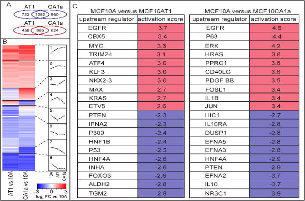
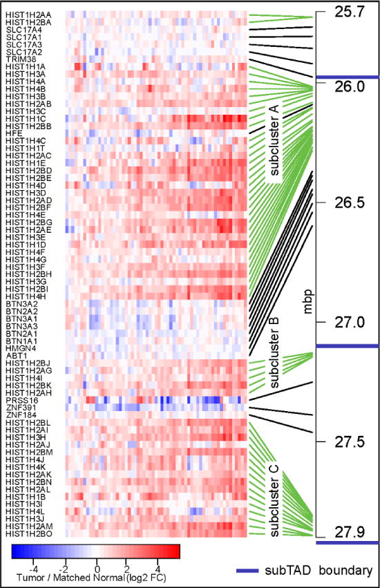
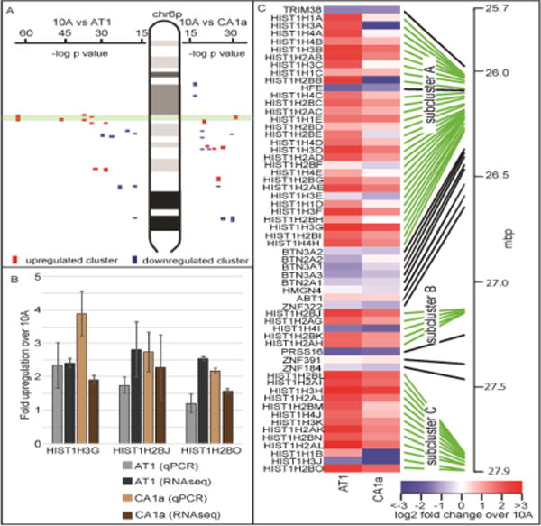
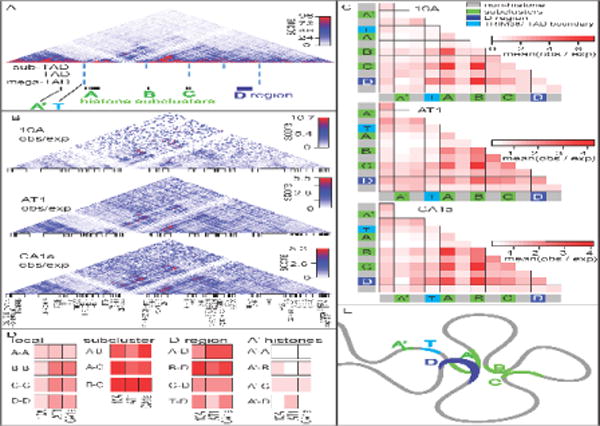
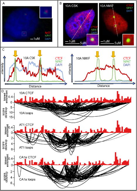
Alterations in nuclear morphology are common in cancer progression. However, the degree to which gross morphological abnormalities translate into compromised higher-order chromatin organization is poorly understood. To explore the functional links between gene expression and chromatin structure in breast cancer, we performed RNA-seq gene expression analysis on the basal breast cancer progression model based on human MCF10A cells. Positional gene enrichment identified the major histone gene cluster at chromosome 6p22 as one of the most significantly upregulated (and not amplified) clusters of genes from the normal-like MCF10A to premalignant MCF10AT1 and metastatic MCF10CA1a cells. This cluster is subdivided into three sub-clusters of histone genes that are organized into hierarchical topologically associating domains (TADs). Interestingly, the sub-clusters of histone genes are located at TAD boundaries and interact more frequently with each other than the regions in-between them, suggesting that the histone sub-clusters form an active chromatin hub. The anchor sites of loops within this hub are occupied by CTCF, a known chromatin organizer. These histone genes are transcribed and processed at a specific sub-nuclear microenvironment termed the major histone locus body (HLB). While the overall chromatin structure of the major HLB is maintained across breast cancer progression, we detected alterations in its structure that may relate to gene expression. Importantly, breast tumor specimens also exhibit a coordinate pattern of upregulation across the major histone gene cluster. Our results provide a novel insight into the connection between the higher-order chromatin organization of the major HLB and its regulation during breast cancer progression.
核形态改变在癌症进展中很常见。然而,大体形态异常转化为高阶染色质组织受损的程度却知之甚少。为了探究乳腺癌中基因表达与染色质结构之间的功能联系,我们基于人MCF10A细胞对基底样乳腺癌进展模型进行了RNA测序基因表达分析。位置基因富集分析确定,位于6号染色体p22区域的主要组蛋白基因簇是从正常样MCF10A细胞到癌前MCF10AT1细胞以及转移性MCF10CA1a细胞中上调最为显著(且未扩增)的基因簇之一。该基因簇被细分为三个组蛋白基因亚簇,它们被组织成层次化的拓扑相关结构域(TAD)。有趣的是,组蛋白基因亚簇位于TAD边界,彼此之间的相互作用比它们之间的区域更为频繁,这表明组蛋白亚簇形成了一个活跃的染色质中心。这个中心内环状结构的锚定位点被CTCF(一种已知的染色质组织者)占据。这些组蛋白基因在一个特定的亚核微环境中进行转录和加工,这个微环境被称为主要组蛋白基因座体(HLB)。虽然在乳腺癌进展过程中主要HLB的整体染色质结构得以维持,但我们检测到其结构的改变可能与基因表达有关。重要的是,乳腺肿瘤标本在主要组蛋白基因簇上也呈现出上调的协同模式。我们的研究结果为乳腺癌进展过程中主要HLB的高阶染色质组织与其调控之间的联系提供了新的见解。