Prashant Kumar, Bhattacharyya Tulsi Das, Frank Herman, Ram Prema
Department of Orthopaedics, Trauma Centre, Institute of Medical Sciences, Banaras Hindu University, Varanasi, Uttar Pradesh, India.
Department of Orthopaedics, Gauhati Medical College & Hospital, Guwahati, Assam, India.
J Orthop Case Rep. 2016 Nov-Dec;6(5):3-6. doi: 10.13107/jocr.2250-0685.604.
Giant cell tumor (GCT) or osteoclastoma is an osteolytic, mostly benign but locally aggressive tumor occurring in young adults at the epiphysis. Area of predilection is mainly long bones (85-90%). 4% of GCT are also found in iliac bone, spine and only 2% in hand (of which GCT phalanges are more common than metacarpal). GCT of metatarsal is a very rare occurrence with very few cases being reported so far. We report a case of GCT 1 metatarsal in a 40-year-old male which is a very rare entity. We shall discuss the clinical features, pathological and radiological hallmarks, and the various treatment modalities of such lesion.
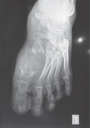
A 40-year-old male presented with complain of swelling over the dorsum of left foot for the duration of 2 years and pain in that foot for 4 months. Swelling was insidious in onset and has progressively increased in size. Pain was mild to moderate in intensity, dull aching and continuous. On examination, there was a localized ovoid shaped swelling 7 by 4 cm over the dorsum of the left foot opposing 1 and 2 metatarsal area with well-defined margins, tender on deep palpation, hard in consistency and the overlying skin was free. Radiographs revealed an expansile osteolytic lesion of entire 1 metatarsal involving the articular surface of tarsometatarsal joint and metatarsophalangeal joint with impingement on 1 metatarsal and cortical thinning. The classical "soap bubble appearance" was also present. Fine needle aspiration cytology was done to confirm our diagnosis of GCT. According to Campanacci ., the tumor was histologically graded as Grade II tumor. A reconstructive surgery with fusion of the Cuneiform metatarsa and metatarsophalangeal joint was planned. The tumor was carefully removed with a cuff of normal tissue and the proximal and distal joints were inspected. There was no articular cartilage of the Cuneiform metatarsa joint. A fibular graft was taken and was inserted into the troughs created in medial cuineform and proximal phalanx and fixed with K-wire, both proximally and distally. The patient was given a below knee cast for three months postoperatively. Full weight bearing was started after 3 months. After 9 months of follow-up, the graft was well taken up and there were no signs of recurrence both clinically and radiologically.
Local resection of the involved metatarsal with autograft or allograft replacement is the preferred surgical treatment for several reasons. First, no correlation has been found between the grade of GCT and the rate of recurrence. Therefore, all giant tumors of foot should be considered locally aggressive. In addition curettage with or without bone grafts has resulted in recurrence rates of about 90%. Thus curettage is an unacceptable form of treatment. Second, although amputation may prevent recurrence, it is cosmetically deforming and decreases the function of the foot.
骨巨细胞瘤(GCT)或破骨细胞瘤是一种溶骨性肿瘤,大多为良性,但具有局部侵袭性,好发于年轻成人的骨骺部位。好发部位主要是长骨(85 - 90%)。4%的骨巨细胞瘤发生于髂骨、脊柱,仅2%发生于手部(其中指骨的骨巨细胞瘤比掌骨更常见)。跖骨骨巨细胞瘤非常罕见,迄今为止报道的病例极少。我们报告一例40岁男性的第一跖骨骨巨细胞瘤,这是一种非常罕见的病例。我们将讨论该病变的临床特征、病理和影像学特点以及各种治疗方式。
一名40岁男性,主诉左脚背肿胀2年,该足部疼痛4个月。肿胀起病隐匿,且逐渐增大。疼痛程度为轻至中度,呈钝痛且持续存在。检查发现,左脚背第一和第二跖骨区域相对处有一个7×4厘米的局限性椭圆形肿胀,边界清晰,深触诊时有压痛,质地硬,皮肤无粘连。X线片显示整个第一跖骨有一个膨胀性溶骨性病变,累及跗跖关节和跖趾关节的关节面,压迫第一跖骨,皮质变薄。还呈现出典型的“肥皂泡样外观”。进行了细针穿刺细胞学检查以确诊骨巨细胞瘤。根据坎帕纳奇分级,该肿瘤组织学分级为二级肿瘤。计划进行楔骨 - 跖骨和跖趾关节融合的重建手术。小心地切除肿瘤并带有一圈正常组织,检查近端和远端关节。楔骨 - 跖骨关节没有关节软骨。取一块腓骨移植骨,插入内侧楔骨和近端指骨所制备的骨槽中,并用克氏针在近端和远端固定。术后给患者使用膝下石膏固定三个月。3个月后开始完全负重。随访9个月后,移植骨愈合良好,临床和影像学均无复发迹象。
因多种原因,对受累跖骨进行局部切除并自体或异体骨移植置换是首选的手术治疗方法。首先,未发现骨巨细胞瘤的分级与复发率之间存在相关性。因此,所有足部的巨大肿瘤都应被视为具有局部侵袭性。此外,刮除术无论是否植骨,复发率约为90%。因此,刮除术是不可接受的治疗方式。其次,虽然截肢可防止复发,但会造成外观畸形并降低足部功能。


