Guo Li, Wang Liang, Wang Li, Yun-Peng Shi, Zhou Jing-Jing, Zhao Zongmao, Li De-Pei
Department of Neurosurgery, The Second Hospital of Hebei Medical University, Shijiazhuang, Hebei 050000, China.
Department of Pharmacy, The Fourth Hospital of Hebei Medical University, Shijiazhuang, Hebei 050011, China.
Stem Cells Int. 2017;2017:1651325. doi: 10.1155/2017/1651325. Epub 2017 Apr 20.
. Human umbilical cord mesenchymal stem cells (hUC-MSCs) potentially differentiate to various types of cells including neuron-like cells. The natural polyphenol resveratrol benefits patients with many diseases including ischemic brain injury. We hypothesize that resveratrol induces differentiation of hUC-MSCs into neuron-like cells. . Flow cytometry was used to determine the surface antigens in different stage of hUC-MSCs (P2, P5, and P10). Nestin, neuron-specific enolase (NSE), and glial fibrillary acidic protein (GFAP) were detected by immunocytochemistry, Western blotting, and real time RT-PCT. The cultured hUC-MSCs were treated with resveratrol at different concentrations (0, 7.5, 15.0, and 30.0 mg/L). Nestin, GFAP, and NSE protein and mRNA were measured at posttreatment time points of 2 h, 4 h, 6 h, 12 h, and 24 h. . Neuron-like cells were found in hUC-MSCs treated by resveratrol at concentrations of 15.0 and 30.0 mg/L, but not in hUC-MSCs treated with vehicle and 7.5 mg/L resveratrol. Furthermore, immunocytochemical staining revealed that nestin and NSE immunoreactivities were positive in resveratrol-treated hUC-MSCs at concentrations of 15.0 and 30.0 mg/L. Resveratrol treatment significantly increased nestin and NSE protein and mRNA levels 4 h after the treatment. However, resveratrol treatment did not change GFAP immunoreactivities and protein and mRNA expression levels in cultured hUC-MSCs. . Taken together, resveratrol treatment induces a differentiation of hUC-MSCs into neuron-like cells at relatively high concentrations.
人脐带间充质干细胞(hUC-MSCs)具有向包括神经元样细胞在内的多种细胞类型分化的潜力。天然多酚白藜芦醇对包括缺血性脑损伤在内的多种疾病患者有益。我们假设白藜芦醇可诱导hUC-MSCs分化为神经元样细胞。
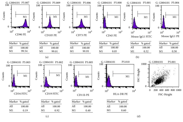
采用流式细胞术检测hUC-MSCs不同阶段(P2、P5和P10)的表面抗原。通过免疫细胞化学、蛋白质印迹法和实时RT-PCR检测巢蛋白、神经元特异性烯醇化酶(NSE)和胶质纤维酸性蛋白(GFAP)。用不同浓度(0、7.5、15.0和30.0mg/L)的白藜芦醇处理培养的hUC-MSCs。在处理后2小时、4小时、6小时、12小时和24小时的时间点测量巢蛋白、GFAP和NSE的蛋白质和mRNA。
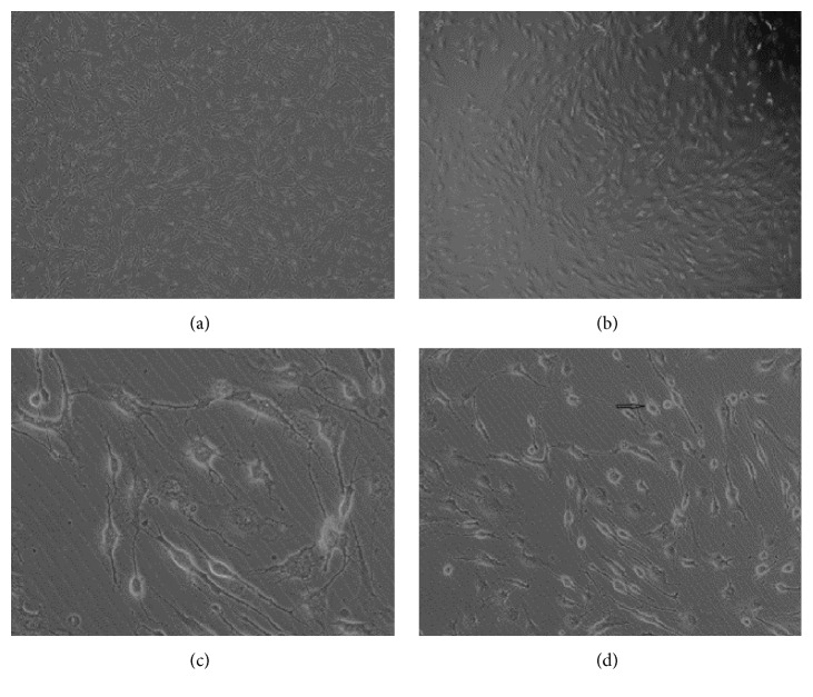
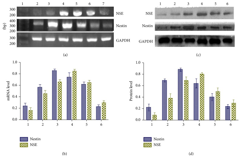
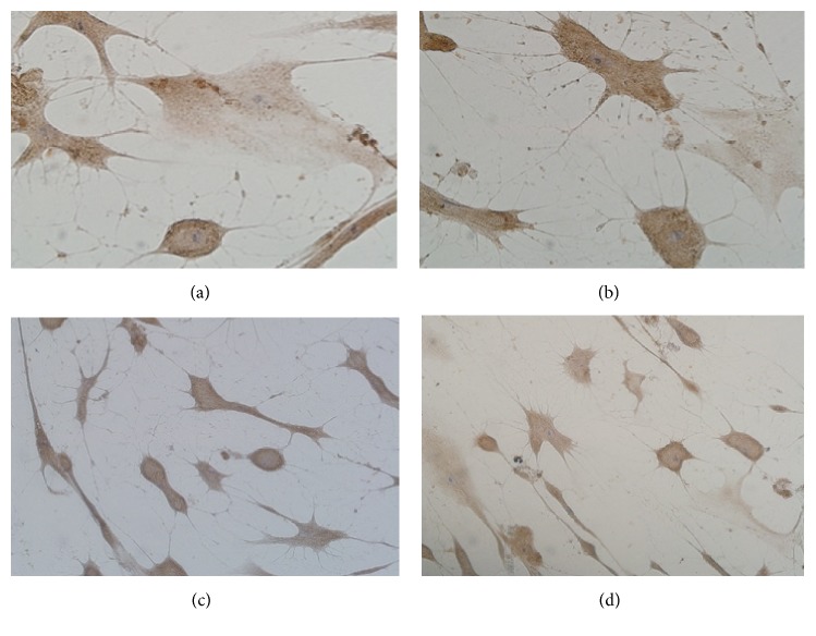
在浓度为15.0和30.0mg/L的白藜芦醇处理的hUC-MSCs中发现了神经元样细胞,但在载体处理和7.5mg/L白藜芦醇处理的hUC-MSCs中未发现。此外,免疫细胞化学染色显示,在浓度为15.0和30.0mg/L的白藜芦醇处理的hUC-MSCs中,巢蛋白和NSE免疫反应呈阳性。白藜芦醇处理后4小时显著增加了巢蛋白和NSE的蛋白质和mRNA水平。然而,白藜芦醇处理并未改变培养的hUC-MSCs中GFAP的免疫反应性以及蛋白质和mRNA表达水平。
综上所述,白藜芦醇处理可诱导较高浓度的hUC-MSCs分化为神经元样细胞。