Ramezanali Fatemeh, Aryanezhad Sasan, Mohammadian Fatemeh, Dibaji Fatemeh, Kharazifard Mohammad Javad
Department of Endodontics, Dental School, International Campus of Tehran University of Medical Sciences, Tehran, Iran.
General Dentist, Tehran, Iran.
Iran Endod J. 2017 Spring;12(2):211-215. doi: 10.22037/iej.2017.41.
This study compared the coronal microleakage of mineral trioxide aggregate (MTA), calcium-enriched mixture (CEM) cement and Biodentine as intra-orifice barriers.
The study was conducted on 76 extracted single-canal human teeth. Their root canals were prepared using ProTaper rotary files and filled with gutta percha and AH-26 sealer using lateral condensation technique. Coronal 3 mm of the gutta percha was removed from the root canals and replaced randomly with MTA, CEM cement or Biodentine in the three experimental groups (=22). A positive and a negative control group were also included (=5). The entire root surfaces of all teeth were covered with two layers of nail varnish in such a way that only the access openings were not coated. In the negative control group, the access opening was also coated with nail varnish. All teeth were immersed in India ink and after clearing, the samples were evaluated under a stereomicroscope under ×10 magnification to assess the degree of dye penetration. The data were analyzed using the Kruskal-Wallis test. The level of significance was set at 0.05.
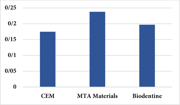
The negative control group showed no leakage while the positive control group showed significantly higher microleakage than the test groups (>0.05). CEM cement had the lowest (0.175±0.068 mm) and MTA showed the highest dye penetration (0.238±0.159 mm) among the experimental groups; although these differences were not statistically significant (=0.313).
CEM cement exhibited the least microleakage as an intra-orifice barrier in endodontically treated teeth.
本研究比较了矿物三氧化物凝聚体(MTA)、富钙混合物(CEM)水泥和生物陶瓷作为根管口屏障时的冠部微渗漏情况。
本研究对76颗拔除的单根管人牙进行。使用ProTaper旋转锉对根管进行预备,采用侧向加压技术用牙胶和AH - 26封闭剂进行充填。从根管中去除冠部3mm的牙胶,在三个实验组(每组n = 22)中随机用MTA、CEM水泥或生物陶瓷进行替换。还设置了一个阳性对照组和一个阴性对照组(每组n = 5)。所有牙齿的整个根面均用两层指甲油覆盖,仅使根管口未被覆盖。在阴性对照组中,根管口也用指甲油覆盖。所有牙齿均浸泡在印度墨水中,清洗后,在体视显微镜下以×10放大倍数评估样本,以评估染料渗透程度。使用Kruskal - Wallis检验分析数据。显著性水平设定为0.05。
阴性对照组无渗漏,而阳性对照组的微渗漏显著高于试验组(P>0.05)。在实验组中,CEM水泥的染料渗透最低(0.175±0.068mm),MTA的染料渗透最高(0.238±0.159mm);尽管这些差异无统计学意义(P = 0.313)。
在根管治疗后的牙齿中,CEM水泥作为根管口屏障时微渗漏最少。