• 文献检索
  • 文档翻译
  • 深度研究
  • 学术资讯
  • Suppr Zotero 插件Zotero 插件
  • 邀请有礼
  • 套餐&价格
  • 历史记录
应用&插件
Suppr Zotero 插件Zotero 插件浏览器插件Mac 客户端Windows 客户端微信小程序
定价
高级版会员购买积分包购买API积分包
服务
文献检索文档翻译深度研究API 文档MCP 服务
关于我们
关于 Suppr公司介绍联系我们用户协议隐私条款
关注我们

Suppr 超能文献

核心技术专利:CN118964589B侵权必究
粤ICP备2023148730 号-1Suppr @ 2026

文献检索

告别复杂PubMed语法,用中文像聊天一样搜索,搜遍4000万医学文献。AI智能推荐,让科研检索更轻松。

立即免费搜索

文件翻译

保留排版,准确专业,支持PDF/Word/PPT等文件格式,支持 12+语言互译。

免费翻译文档

深度研究

AI帮你快速写综述,25分钟生成高质量综述,智能提取关键信息,辅助科研写作。

立即免费体验

酷似脑皮质肿瘤的原发性中枢神经系统血管炎:病例报告

Primary Central Nervous System Vasculitis Mimicking a Cortical Brain Tumor: A Case Report.

作者信息

Lee Joo-Seok, Jung Tae-Young, Lee Kyung-Hwa, Kim Seul-Kee

机构信息

Department of Neurosurgery, Kwangju Christian Hospital, Gwangju, Korea.

Department of Neurosurgery, Chonnam National University Medical School, Chonnam National University Hwasun Hospital, Hwasun, Korea.

出版信息

Brain Tumor Res Treat. 2017 Apr;5(1):30-33. doi: 10.14791/btrt.2017.5.1.30. Epub 2017 Apr 30.

DOI:10.14791/btrt.2017.5.1.30
PMID:28516076
原文链接:https://pmc.ncbi.nlm.nih.gov/articles/PMC5433948/
Abstract

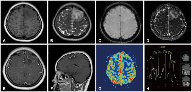
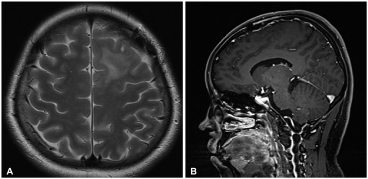
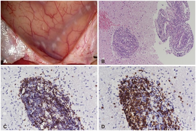
We report a case of primary central nervous system vasculitis (PCNSV) mimicking a cortical brain tumor. A 25-year-old woman presented with a 2-week history of headache and transient right hemiparesis. Brain magnetic resonance imaging (MRI) revealed a cortical-involving lesion on the left frontal lobe. The 6-cm sized lesion showed low signal intensity on T1-weighted images and high signal intensity on T2-weighted images. The lesion had continual linear enhancement on the subcortical white matter and leptomeninges. There was no evidence of hemorrhage on susceptibility-weighted images and no diffusion restriction on diffusion-weighted images. The regional cerebral blood volume was decreased on the MR perfusion images, and spectroscopy showed increased lactate and lipid peaks. The symptoms were aggravated by fever and seizures. Biopsy was performed to rule out tumorous or inflammatory lesions. Pathologically, lymphocytes were infiltrated on the vessels, and the arachnoid membrane was thickened with inflammatory cells. The patient did not have any underlying diseases, including immune disorders. After high-dose steroid administration, her symptoms improved. Two months later, brain MRI showed a reduction in the infiltration of the T2 hyperintensity lesion with subtle subcortical enhancement. We present a case of PCNSV involving the left frontal lobe, showing vasogenic edema, mass effect, and subcortical linear contrast enhancement without hemorrhage or infarction.

摘要

我们报告一例疑似皮质脑肿瘤的原发性中枢神经系统血管炎(PCNSV)病例。一名25岁女性,有2周头痛及短暂性右半身轻瘫病史。脑部磁共振成像(MRI)显示左额叶有一累及皮质的病变。该6厘米大小的病变在T1加权图像上呈低信号强度,在T2加权图像上呈高信号强度。病变在皮质下白质和软脑膜有持续的线性强化。磁敏感加权图像上无出血迹象,扩散加权图像上无扩散受限。MR灌注图像显示局部脑血容量减少,磁共振波谱显示乳酸和脂质峰升高。发热和癫痫发作使症状加重。进行活检以排除肿瘤性或炎性病变。病理检查显示血管有淋巴细胞浸润,蛛网膜增厚并有炎性细胞。该患者无任何基础疾病,包括免疫紊乱。大剂量类固醇治疗后,她的症状有所改善。两个月后,脑部MRI显示T2高信号病变的浸润减少,皮质下有轻微强化。我们报告一例累及左额叶的PCNSV病例,表现为血管源性水肿、占位效应及皮质下线性对比增强,无出血或梗死。

https://cdn.ncbi.nlm.nih.gov/pmc/blobs/2b77/5433948/aba3f2f28d4a/btrt-5-30-g003.jpg
https://cdn.ncbi.nlm.nih.gov/pmc/blobs/2b77/5433948/4f720ce1acb3/btrt-5-30-g001.jpg
https://cdn.ncbi.nlm.nih.gov/pmc/blobs/2b77/5433948/954572d3d82a/btrt-5-30-g002.jpg
https://cdn.ncbi.nlm.nih.gov/pmc/blobs/2b77/5433948/aba3f2f28d4a/btrt-5-30-g003.jpg
https://cdn.ncbi.nlm.nih.gov/pmc/blobs/2b77/5433948/4f720ce1acb3/btrt-5-30-g001.jpg
https://cdn.ncbi.nlm.nih.gov/pmc/blobs/2b77/5433948/954572d3d82a/btrt-5-30-g002.jpg
https://cdn.ncbi.nlm.nih.gov/pmc/blobs/2b77/5433948/aba3f2f28d4a/btrt-5-30-g003.jpg

相似文献

1
Primary Central Nervous System Vasculitis Mimicking a Cortical Brain Tumor: A Case Report.酷似脑皮质肿瘤的原发性中枢神经系统血管炎:病例报告
Brain Tumor Res Treat. 2017 Apr;5(1):30-33. doi: 10.14791/btrt.2017.5.1.30. Epub 2017 Apr 30.
2
Spontaneously changing MRI findings of primary central nervous system vasculitis: A case report.原发性中枢神经系统血管炎的 MRI 表现自发性改变:一例报告。
J Clin Neurosci. 2021 Jan;83:125-127. doi: 10.1016/j.jocn.2020.11.012. Epub 2020 Nov 24.
3
[A central nervous system lupus showing peculiar findings on cranial magnetic resonance imaging (MRI)].[一例在头颅磁共振成像(MRI)上显示特殊表现的中枢神经系统狼疮]
Rinsho Shinkeigaku. 2003 Jul;43(7):409-16.
4
[Positron emission tomographic evaluation for frontal hypertrophic cranial pachymeningitis using 11C-methyl-L-methionine].[使用11C-甲基-L-蛋氨酸对额部肥厚性硬脑膜炎进行正电子发射断层扫描评估]
No Shinkei Geka. 1996 Mar;24(3):287-93.
5
Diffusion-weighted MR of the brain: methodology and clinical application.脑部扩散加权磁共振成像:方法与临床应用
Radiol Med. 2005 Mar;109(3):155-97.
6
[A case of cerebral amyloid angiopathy with reversible white matter lesions and multiple cerebral microbleeds].一例伴有可逆性白质病变和多发性脑微出血的脑淀粉样血管病
Rinsho Shinkeigaku. 2012;52(2):90-5. doi: 10.5692/clinicalneurol.52.90.
7
Diffusion-weighted magnetic resonance images in a patient with neuropsychiatric lupus.一名神经精神性狼疮患者的磁共振扩散加权成像
J Clin Neurosci. 2007 Mar;14(3):273-5. doi: 10.1016/j.jocn.2005.12.050.
8
Case 259: Primary Central Nervous System Lymphomatoid Granulomatosis Mimicking Chronic Lymphocytic Inflammation with Pontine Perivascular Enhancement Responsive to Steroids (CLIPPERS).病例 259:原发性中枢神经系统淋巴瘤样肉芽肿病,类似伴有桥脑血管周围强化的慢性淋巴细胞性炎症对类固醇反应(CLIPPERS)。
Radiology. 2018 Nov;289(2):572-577. doi: 10.1148/radiol.2018161475.
9
[Longitudinally Extensive Transverse Myelitis Caused by Granulomatous Primary Central Nervous System Vasculitis with Characteristic Brain Magnetic Resonance Image: A Case Report].[肉芽肿性原发性中枢神经系统血管炎伴特征性脑磁共振成像导致的纵向广泛横贯性脊髓炎:一例报告]
Brain Nerve. 2021 Aug;73(8):931-937. doi: 10.11477/mf.1416201863.
10
[Early diagnostic significance and dynamic pattern of DWI compared with conventional MRI in newborns with neonatal cerebral infarction].[新生儿脑梗死患儿中磁共振扩散加权成像(DWI)与传统磁共振成像(MRI)对比的早期诊断意义及动态变化模式]
Zhonghua Er Ke Za Zhi. 2007 May;45(5):360-4.

引用本文的文献

1
Clinical and diagnostic characteristics of Hashimoto's encephalopathy: a single-center, retrospective study.桥本脑病的临床与诊断特征:一项单中心回顾性研究
Acta Neurol Belg. 2024 Dec;124(6):1839-1846. doi: 10.1007/s13760-024-02520-1. Epub 2024 Jun 11.
2
Differential Diagnosis of Tumor-like Brain Lesions.脑肿瘤样病变的鉴别诊断
Neurol Clin Pract. 2023 Oct;13(5):e200182. doi: 10.1212/CPJ.0000000000200182. Epub 2023 Aug 30.
3
Anaplastic pleomorphic xanthoastrocytoma misdiagnosed as cerebral sparganosis-identification of the "mirror image".

本文引用的文献

1
[Treatment for central nervous system vasculitis].[中枢神经系统血管炎的治疗]
Brain Nerve. 2015 Mar;67(3):287-93. doi: 10.11477/mf.1416200133.
2
Cerebral tumor or pseudotumor?脑瘤还是假瘤?
Diagn Interv Imaging. 2014 Oct;95(10):906-16. doi: 10.1016/j.diii.2014.08.004. Epub 2014 Sep 26.
3
Adult primary central nervous system vasculitis.成人原发性中枢神经系统血管炎。
误诊为脑裂头蚴病的间变性多形性黄色星形细胞瘤——“镜像”的识别
Quant Imaging Med Surg. 2021 Oct;11(10):4479-4487. doi: 10.21037/qims-20-1398.
4
Tumefactive Primary Central Nervous System Vasculitis: Imaging Findings of a Rare and Underrecognized Neuroinflammatory Disease.肿块样原发性中枢神经系统血管炎:一种罕见且认识不足的神经炎症性疾病的影像学表现。
AJNR Am J Neuroradiol. 2020 Nov;41(11):2075-2081. doi: 10.3174/ajnr.A6736. Epub 2020 Sep 3.
5
"When should primary angiitis of the central nervous system (PACNS) be suspected?": literature review and proposal of a preliminary screening algorithm.“何时应怀疑中枢神经系统原发性血管炎(PACNS)?”:文献综述及初步筛查算法建议
Neurol Sci. 2020 Nov;41(11):3135-3148. doi: 10.1007/s10072-020-04583-3. Epub 2020 Aug 10.
6
A Case of Primary Central Nervous System Vasculitis That Worsened Despite Early Corticosteroid Therapy.一例尽管早期使用皮质类固醇治疗仍恶化的原发性中枢神经系统血管炎病例。
Brain Tumor Res Treat. 2019 Oct;7(2):164-167. doi: 10.14791/btrt.2019.7.e37.
7
Clinical value of magnetic resonance imaging in identifying multiple cerebral gliomas from primary central nervous system lymphoma.磁共振成像在鉴别原发性中枢神经系统淋巴瘤与多发性脑胶质瘤中的临床价值
Oncol Lett. 2019 Jul;18(1):593-598. doi: 10.3892/ol.2019.10352. Epub 2019 May 13.
Lancet. 2012 Aug 25;380(9843):767-77. doi: 10.1016/S0140-6736(12)60069-5. Epub 2012 May 9.
4
Primary angiitis of the central nervous system mimicking tumor-like lesion--case report.酷似肿瘤样病变的中枢神经系统原发性血管炎——病例报告
Neurol Med Chir (Tokyo). 2011;51(1):56-9. doi: 10.2176/nmc.51.56.
5
Diagnosis and treatment of cerebral vasculitis.脑血管炎的诊断与治疗。
Ther Adv Neurol Disord. 2010 Jan;3(1):29-42. doi: 10.1177/1756285609347123.
6
Tumor-like lesions of the brain.脑内肿瘤样病变。
Cancer Imaging. 2009 Oct 2;9 Spec No A(Special issue A):S10-3. doi: 10.1102/1470-7330.2009.9003.
7
Primary angiitis of the central nervous system.中枢神经系统原发性血管炎
Arch Neurol. 2009 Jun;66(6):704-9. doi: 10.1001/archneurol.2009.76.
8
[Primary central nervous system vasculitis imitating a brain tumour--a case report].[酷似脑肿瘤的原发性中枢神经系统血管炎——病例报告]
Neurol Neurochir Pol. 2008 Jul-Aug;42(4):358-61.
9
Tumour-like mass lesion: an under-recognised presentation of primary angiitis of the central nervous system.肿瘤样肿块病变:一种未被充分认识的中枢神经系统原发性血管炎表现。
Ann Rheum Dis. 2008 Dec;67(12):1732-5. doi: 10.1136/ard.2008.096800. Epub 2008 Jul 14.
10
Primary central nervous system vasculitis: analysis of 101 patients.原发性中枢神经系统血管炎:101例患者分析
Ann Neurol. 2007 Nov;62(5):442-51. doi: 10.1002/ana.21226.