• 文献检索
  • 文档翻译
  • 深度研究
  • 学术资讯
  • Suppr Zotero 插件Zotero 插件
  • 邀请有礼
  • 套餐&价格
  • 历史记录
应用&插件
Suppr Zotero 插件Zotero 插件浏览器插件Mac 客户端Windows 客户端微信小程序
定价
高级版会员购买积分包购买API积分包
服务
文献检索文档翻译深度研究API 文档MCP 服务
关于我们
关于 Suppr公司介绍联系我们用户协议隐私条款
关注我们

Suppr 超能文献

核心技术专利:CN118964589B侵权必究
粤ICP备2023148730 号-1Suppr @ 2026

文献检索

告别复杂PubMed语法,用中文像聊天一样搜索,搜遍4000万医学文献。AI智能推荐,让科研检索更轻松。

立即免费搜索

文件翻译

保留排版,准确专业,支持PDF/Word/PPT等文件格式,支持 12+语言互译。

免费翻译文档

深度研究

AI帮你快速写综述,25分钟生成高质量综述,智能提取关键信息,辅助科研写作。

立即免费体验

利用超声检查评估甲状腺乳头状癌的包膜侵犯或包膜外扩展与淋巴结转移之间的关系。

Using Ultrasonography to Evaluate the Relationship between Capsular Invasion or Extracapsular Extension and Lymph Node Metastasis in Papillary Thyroid Carcinomas.

作者信息

Jiao Wei-Ping, Zhang Lei

机构信息

Department of Ultrasound, Beijing Xuanwu Hospital, Capital Medical University, Beijing 100053, China.

Department of Cardiology, Beijing Xuanwu Hospital, Capital Medical University, Beijing 100053, China.

出版信息

Chin Med J (Engl). 2017 Jun 5;130(11):1309-1313. doi: 10.4103/0366-6999.206339.

DOI:10.4103/0366-6999.206339
PMID:28524830
原文链接:https://pmc.ncbi.nlm.nih.gov/articles/PMC5455040/
Abstract

BACKGROUND

Previous studies have demonstrated that ultrasonography is the recommended imaging modality for preoperative staging of papillary thyroid carcinomas (PTCs). However, only a few studies have kept watch on preoperative evaluation of capsular invasion (CI) or extracapsular extension (ECE) and cervical lymph node metastasis using preoperative ultrasonography. This study aimed to investigate the relationship between the CI or ECE and the cervical lymph node metastasis in PTCs using preoperative ultrasonography and postoperative pathology in Chinese patients.

METHODS

The data of preoperative ultrasonography and postoperative pathology of 166 patients who had a definitive diagnosis of PTCs from October 2011 to July 2014 at Xuanwu Hospital, Beijing were collected and reviewed. Preoperative ultrasonic parameters of thyroid nodules were compared with those of postoperative pathological diagnoses. All the patients were divided into bilateral PTCs group (n = 42, 78 nodules) and unilateral PTCs group (n = 124, 124 nodules), and the data of the nodule sizes, CI or ECE, and cervical lymph node metastasis by preoperative ultrasonography were compared between two groups.

RESULTS

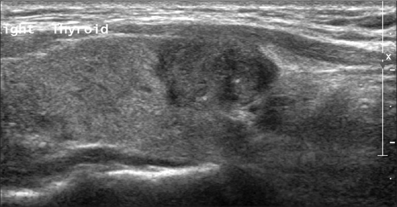
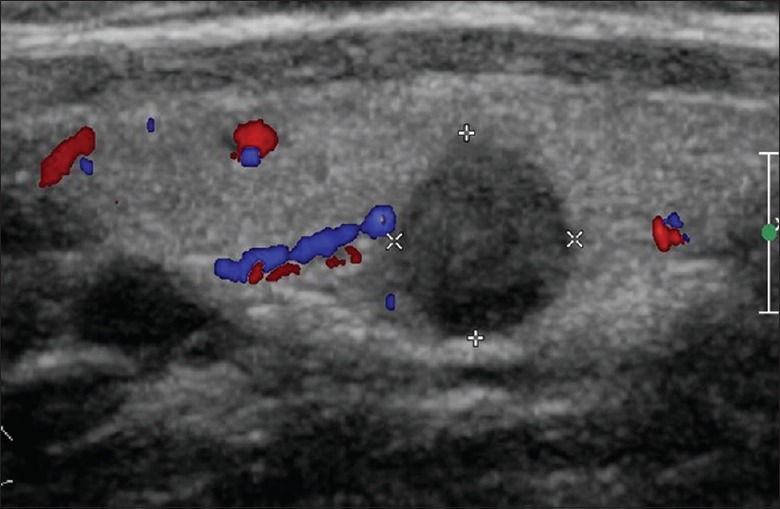
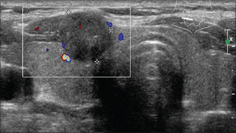
A total of 202 nodules of 166 patients which were confirmed by preoperative ultrasonography and postoperative pathology were analyzed. Hypoechogenicity (n = 201, 99.5%) and irregular margins (n = 167, 82.7%) were the main ultrasonic characteristics of PTCs. A significant moderate agreement between preoperative ultrasonic examination and postoperative pathology for CI or ECE (κ = 0.622, P< 0.001) was observed. The diagnostic sensitivity was 92.0%, and specificity was 71.1%. In bilateral PTCs group, 81.0% had CI or ECE, and 61.9% had cervical lymph node metastasis. In unilateral PTCs group, 76.6% had CI or ECE, and 58.1% had cervical lymph node metastasis. There were no significant differences in the incidence of CI or ECE and cervical lymph node metastasis between two groups (all P > 0.05).

CONCLUSIONS

Ultrasonography was proved to be a valuable method for preoperative diagnosis of PTCs. Hypoechogenicity and irregular margins were strongly associated with PTCs. CI or ECE in unilateral PTCs strongly implied the cervical lymph node metastasis. Therefore, the cervical lymph nodes should be carefully examined by ultrasonography in patients with PTCs.

摘要

背景

既往研究表明,超声检查是推荐用于甲状腺乳头状癌(PTC)术前分期的影像学检查方法。然而,仅有少数研究关注术前超声检查对甲状腺被膜侵犯(CI)或包膜外扩展(ECE)及颈部淋巴结转移的评估。本研究旨在通过术前超声检查及术后病理,探讨中国PTC患者中CI或ECE与颈部淋巴结转移之间的关系。

方法

收集并回顾2011年10月至2014年7月在北京宣武医院确诊为PTC的166例患者的术前超声检查及术后病理数据。将甲状腺结节的术前超声参数与术后病理诊断结果进行比较。所有患者分为双侧PTC组(n = 42,78个结节)和单侧PTC组(n = 124,124个结节),比较两组术前超声检查的结节大小、CI或ECE及颈部淋巴结转移情况。

结果

对166例患者经术前超声检查及术后病理证实的202个结节进行分析。低回声(n = 201,99.5%)和边界不规则(n = 167,82.7%)是PTC的主要超声特征。术前超声检查与术后病理对CI或ECE的诊断有显著的中度一致性(κ = 0.622,P < 0.001)。诊断敏感性为92.0%,特异性为71.1%。双侧PTC组中,81.0%有CI或ECE,61.9%有颈部淋巴结转移。单侧PTC组中,76.6%有CI或ECE,58.1%有颈部淋巴结转移。两组间CI或ECE及颈部淋巴结转移的发生率无显著差异(均P > 0.05)。

结论

超声检查被证明是PTC术前诊断的一种有价值的方法。低回声和边界不规则与PTC密切相关。单侧PTC中的CI或ECE强烈提示颈部淋巴结转移。因此,PTC患者应通过超声仔细检查颈部淋巴结。

https://cdn.ncbi.nlm.nih.gov/pmc/blobs/82b9/5455040/fa0e8ba2f56a/CMJ-130-1309-g003.jpg
https://cdn.ncbi.nlm.nih.gov/pmc/blobs/82b9/5455040/51a8aa5a6ef6/CMJ-130-1309-g001.jpg
https://cdn.ncbi.nlm.nih.gov/pmc/blobs/82b9/5455040/90d9802e123e/CMJ-130-1309-g002.jpg
https://cdn.ncbi.nlm.nih.gov/pmc/blobs/82b9/5455040/fa0e8ba2f56a/CMJ-130-1309-g003.jpg
https://cdn.ncbi.nlm.nih.gov/pmc/blobs/82b9/5455040/51a8aa5a6ef6/CMJ-130-1309-g001.jpg
https://cdn.ncbi.nlm.nih.gov/pmc/blobs/82b9/5455040/90d9802e123e/CMJ-130-1309-g002.jpg
https://cdn.ncbi.nlm.nih.gov/pmc/blobs/82b9/5455040/fa0e8ba2f56a/CMJ-130-1309-g003.jpg

相似文献

1
Using Ultrasonography to Evaluate the Relationship between Capsular Invasion or Extracapsular Extension and Lymph Node Metastasis in Papillary Thyroid Carcinomas.利用超声检查评估甲状腺乳头状癌的包膜侵犯或包膜外扩展与淋巴结转移之间的关系。
Chin Med J (Engl). 2017 Jun 5;130(11):1309-1313. doi: 10.4103/0366-6999.206339.
2
Contrast-enhanced ultrasound: a valuable modality for extracapsular extension assessment in papillary thyroid cancer.超声造影:评估甲状腺乳头状癌囊外侵犯的有价值的影像学方法。
Eur Radiol. 2021 Jul;31(7):4568-4575. doi: 10.1007/s00330-020-07516-y. Epub 2021 Jan 7.
3
[Study on the relationship between ultrasonographic features of papillary thyroid carcinoma and central cervical lymph node metastasis].甲状腺乳头状癌超声特征与中央区颈部淋巴结转移关系的研究
Zhonghua Zhong Liu Za Zhi. 2018 Mar 23;40(3):196-200. doi: 10.3760/cma.j.issn.0253-3766.2018.03.007.
4
[Diagnosis of lateral cervical lymph node metastasis of papillary carcinoma of the thyroid by ultrasonography].[超声检查对甲状腺乳头状癌颈侧区淋巴结转移的诊断]
Nihon Jibiinkoka Gakkai Kaiho. 2004 Dec;107(12):1038-44. doi: 10.3950/jibiinkoka.107.1038.
5
[Role of preoperative ultrasonography in evaluating early cervical metastasis in patients with differentiated thyroid carcinoma].[术前超声检查在评估分化型甲状腺癌患者早期颈部转移中的作用]
Zhongguo Yi Xue Ke Xue Yuan Xue Bao. 2006 Aug;28(4):530-3.
6
Cervical lymph node metastases in papillary thyroid cancer: Preoperative staging with ultrasound and/or computed tomography.甲状腺乳头状癌颈淋巴结转移:超声和/或计算机断层扫描的术前分期。
Medicine (Baltimore). 2022 Mar 4;101(9):e28909. doi: 10.1097/MD.0000000000028909.
7
Clinicopathologic Factors and Thyroid Nodule Sonographic Features for Predicting Central Lymph Node Metastasis in Papillary Thyroid Microcarcinoma: A Retrospective Study of 1204 Patients.预测甲状腺微小乳头状癌中央区淋巴结转移的临床病理因素及甲状腺结节超声特征:一项对1204例患者的回顾性研究
J Ultrasound Med. 2016 Nov;35(11):2475-2481. doi: 10.7863/ultra.15.10012. Epub 2016 Oct 25.
8
Lymph node metastasis characteristics of papillary thyroid carcinoma located in the isthmus: A single-center analysis.位于甲状腺峡部的乳头状甲状腺癌的淋巴结转移特征:单中心分析。
Medicine (Baltimore). 2017 Jun;96(24):e7143. doi: 10.1097/MD.0000000000007143.
9
Prediction of thyroid extracapsular extension with cervical lymph node metastases (ECE-LN) by CEUS and BRAF expression in papillary thyroid carcinoma.通过对比增强超声(CEUS)和BRAF表达预测甲状腺乳头状癌伴颈部淋巴结转移的甲状腺包膜外侵犯(ECE-LN)情况
Tumour Biol. 2014 Sep;35(9):8559-64. doi: 10.1007/s13277-014-2119-2. Epub 2014 May 27.
10
Prediction of cervical lymph node metastasis in patients with papillary thyroid cancer using combined conventional ultrasound, strain elastography, and acoustic radiation force impulse (ARFI) elastography.联合应用传统超声、应变弹性成像及声辐射力脉冲(ARFI)弹性成像预测甲状腺乳头状癌患者颈部淋巴结转移
Eur Radiol. 2016 Aug;26(8):2611-22. doi: 10.1007/s00330-015-4088-2. Epub 2015 Nov 11.

引用本文的文献

1
Impact of Ultrasonographic Features Indicative of Malignancy on Tumor Advancement in Thyroid Cancer-A Single-Center Study.超声提示恶性特征对甲状腺癌肿瘤进展的影响——一项单中心研究
Cancers (Basel). 2025 Aug 28;17(17):2822. doi: 10.3390/cancers17172822.
2
Positive preoperative circulating tumor cells level associated with lymph node metastasis in papillary thyroid carcinoma patients with capsular invasion.术前循环肿瘤细胞水平呈阳性与有包膜侵犯的乳头状甲状腺癌患者的淋巴结转移相关。
World J Surg Oncol. 2025 May 14;23(1):190. doi: 10.1186/s12957-025-03842-x.
3
Omniview of three-dimensional ultrasound for prospective evaluation of extrathyroidal extension of differentiated thyroid cancer.

本文引用的文献

1
Evaluation of Diagnostic Efficiency of Ultrasound Features on Malignant Thyroid Nodules in Chinese Patients.超声特征对中国患者甲状腺恶性结节的诊断效能评估
Chin Med J (Engl). 2016 Aug 5;129(15):1784-8. doi: 10.4103/0366-6999.186643.
2
"Onion Skin-liked Sign" in Thyroid Ultrasonography: A Characteristic Feature of Benign Thyroid Nodules.甲状腺超声检查中的“葱皮样征”:良性甲状腺结节的特征性表现
Chin Med J (Engl). 2016 Jul 5;129(13):1533-7. doi: 10.4103/0366-6999.184460.
3
Predictive value of nodal metastases on local recurrence in the management of differentiated thyroid cancer. Retrospective clinical study.
三维超声前瞻性评估分化型甲状腺癌甲状腺外侵犯的全景观察
BMC Med Imaging. 2025 Feb 10;25(1):42. doi: 10.1186/s12880-025-01572-w.
4
Factors influencing extrathyroidal extension of papillary thyroid cancer and evaluation of ultrasonography for its diagnosis: a retrospective analysis.影响甲状腺外侵犯的因素及超声对此的诊断价值:回顾性分析。
Sci Rep. 2023 Oct 26;13(1):18344. doi: 10.1038/s41598-023-45642-x.
5
Nomogram incorporating preoperative clinical and ultrasound indicators to predict aggressiveness of solitary papillary thyroid carcinoma.纳入术前临床和超声指标的列线图,用于预测孤立性甲状腺乳头状癌的侵袭性。
Front Oncol. 2023 Jan 31;13:1009958. doi: 10.3389/fonc.2023.1009958. eCollection 2023.
6
The Diagnostic Performance of Ultrasonography in the Evaluation of Extrathyroidal Extension in Papillary Thyroid Carcinoma: A Systematic Review and Meta-Analysis.超声检查在评估甲状腺乳头状癌甲状腺外侵犯中的诊断性能:系统评价和荟萃分析。
Int J Mol Sci. 2022 Dec 26;24(1):371. doi: 10.3390/ijms24010371.
7
Clinical Value of Ultrasonography and Serum Markers in Preoperative N Staging of Thyroid Cancer.超声检查及血清标志物在甲状腺癌术前 N 分期中的临床价值。
Cells. 2022 Nov 15;11(22):3621. doi: 10.3390/cells11223621.
8
Risk factors for central lymph node metastasis in the cervical region in papillary thyroid carcinoma: a retrospective study.甲状腺乳头状癌颈部中央淋巴结转移的危险因素:一项回顾性研究
World J Surg Oncol. 2021 Apr 30;19(1):138. doi: 10.1186/s12957-021-02247-w.
分化型甲状腺癌治疗中淋巴结转移对局部复发的预测价值。回顾性临床研究。
BMC Surg. 2013;13 Suppl 2(Suppl 2):S3. doi: 10.1186/1471-2482-13-S2-S3. Epub 2013 Oct 8.
4
Preoperative prediction of central lymph node metastasis in thyroid papillary microcarcinoma using clinicopathologic and sonographic features.术前应用临床病理和超声特征预测甲状腺乳头状微小癌中央区淋巴结转移。
World J Surg. 2013 Feb;37(2):385-91. doi: 10.1007/s00268-012-1826-3.
5
Extent of surgery for papillary thyroid cancer: preoperative imaging and role of prophylactic and therapeutic neck dissection.甲状腺乳头状癌的手术范围:术前影像学检查及预防性和治疗性颈清扫术的作用。
Curr Treat Options Oncol. 2012 Mar;13(1):1-10. doi: 10.1007/s11864-011-0175-z.
6
Role of ultrasound, clinical and scintigraphyc parameters to predict malignancy in thyroid nodule.超声、临床及闪烁扫描参数在预测甲状腺结节恶性程度中的作用。
Head Neck Oncol. 2011 Mar 22;3:17. doi: 10.1186/1758-3284-3-17.
7
Management of thyroid nodules and surgery for differentiated thyroid cancer.甲状腺结节的管理和分化型甲状腺癌的手术治疗。
Clin Oncol (R Coll Radiol). 2010 Aug;22(6):405-12. doi: 10.1016/j.clon.2010.03.009. Epub 2010 Apr 8.
8
Clinical and imaging assessment of cervical lymph node metastasis in papillary thyroid carcinomas.甲状腺乳头状癌颈淋巴结转移的临床与影像学评估。
World J Surg. 2010 Jul;34(7):1494-9. doi: 10.1007/s00268-010-0541-1.
9
Papillary thyroid carcinoma on sonography.超声检查下的甲状腺乳头状癌。
Clin Imaging. 2010 Mar-Apr;34(2):121-6. doi: 10.1016/j.clinimag.2009.03.003.
10
Developments in the use of ultrasound for thyroid cancer.超声在甲状腺癌中的应用进展。
Curr Opin Oncol. 2010 Jan;22(1):11-6. doi: 10.1097/CCO.0b013e3283337f16.