Department of Ophthalmology, Margaret Dyson Vision Research Institute, Weill Cornell Medicine, 1300 York Avenue, New York, New York 10065, USA.
Angiocrine Bioscience, Inc., 11575 Sorrento Valley Road, Suite 217, San Diego, California 92121, USA.
Nat Commun. 2017 May 19;8:15374. doi: 10.1038/ncomms15374.
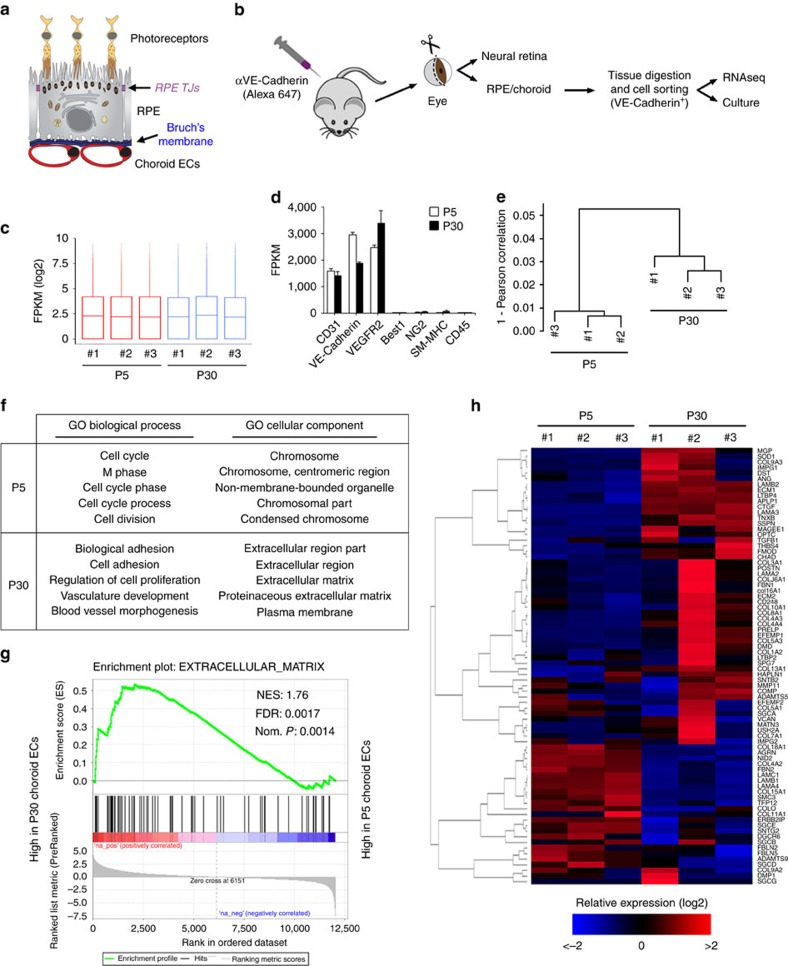
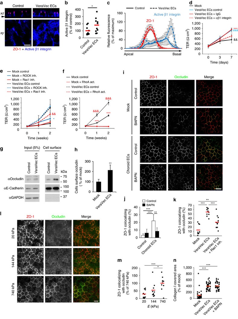
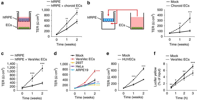
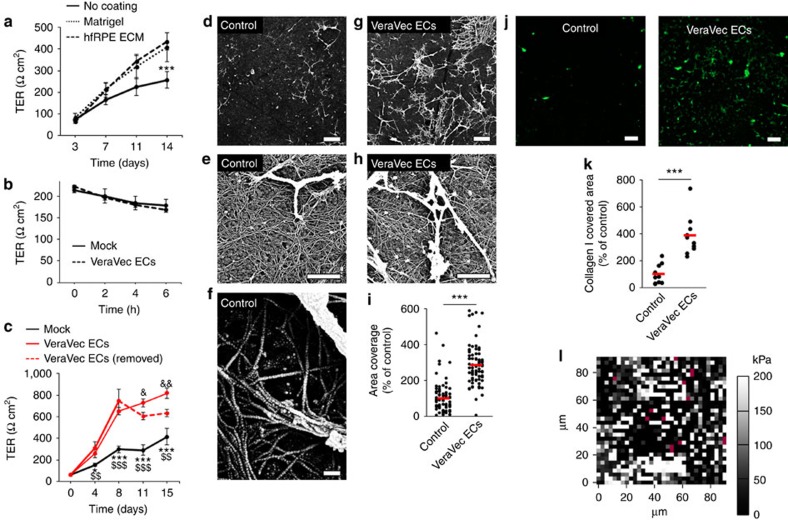
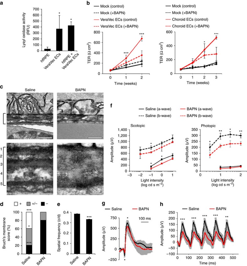
The outer blood-retina barrier is established through the coordinated terminal maturation of the retinal pigment epithelium (RPE), fenestrated choroid endothelial cells (ECs) and Bruch's membrane, a highly organized basement membrane that lies between both cell types. Here we study the contribution of choroid ECs to this process by comparing their gene expression profile before (P5) and after (P30) the critical postnatal period when mice acquire mature visual function. Transcriptome analyses show that expression of extracellular matrix-related genes changes dramatically over this period. Co-culture experiments support the existence of a novel regulatory pathway: ECs secrete factors that remodel RPE basement membrane, and integrin receptors sense these changes triggering Rho GTPase signals that modulate RPE tight junctions and enhance RPE barrier function. We anticipate our results will spawn a search for additional roles of choroid ECs in RPE physiology and disease.
外血视网膜屏障是通过视网膜色素上皮(RPE)、有孔脉络膜内皮细胞(EC)和布鲁赫膜的协调终末成熟来建立的,布鲁赫膜是一种位于这两种细胞类型之间的高度组织化的基底膜。在这里,我们通过比较在小鼠获得成熟视觉功能的关键出生后时期之前(P5)和之后(P30)的脉络膜 EC 的基因表达谱来研究它们对这一过程的贡献。转录组分析表明,在此期间细胞外基质相关基因的表达发生了剧烈变化。共培养实验支持存在一种新的调节途径:EC 分泌重塑 RPE 基底膜的因子,整合素受体感知这些变化,触发 Rho GTPase 信号,调节 RPE 紧密连接并增强 RPE 屏障功能。我们预计我们的结果将引发对脉络膜 EC 在 RPE 生理学和疾病中的其他作用的研究。