Department of Orthopaedic Surgery, Tokyo Medical and Dental University, 1-5-45 Yushima, Bunkyo-ku, Tokyo, 113-8510, Japan.
Department of Advanced Technology in Medicine, Graduate School of Tokyo Medical and Dental University, 1-5-45 Yushima, Bunkyo-ku, Tokyo, 113-8510, Japan.
Sci Rep. 2017 May 19;7(1):2192. doi: 10.1038/s41598-017-02406-8.
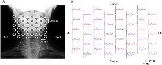
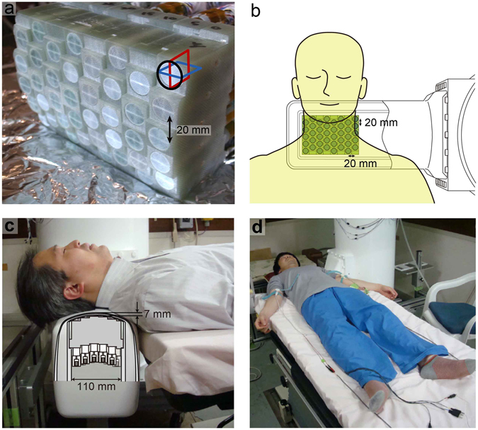
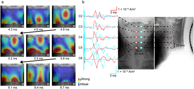
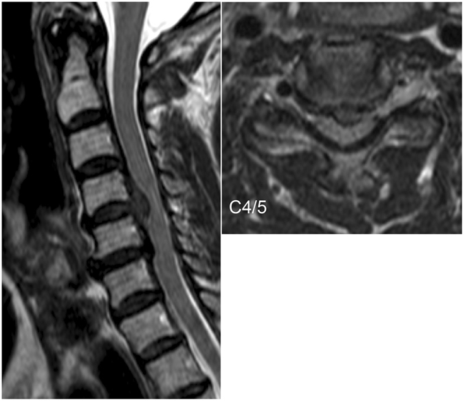
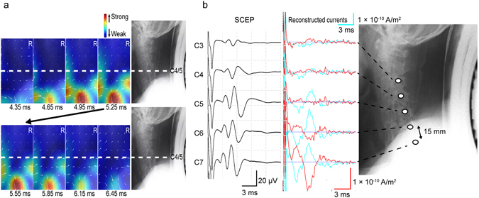
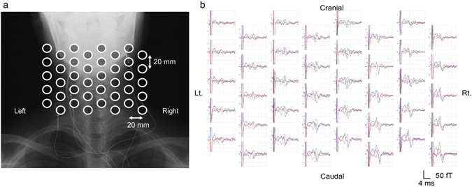
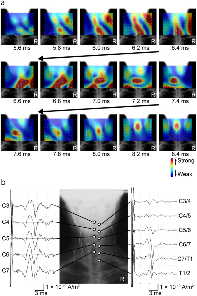
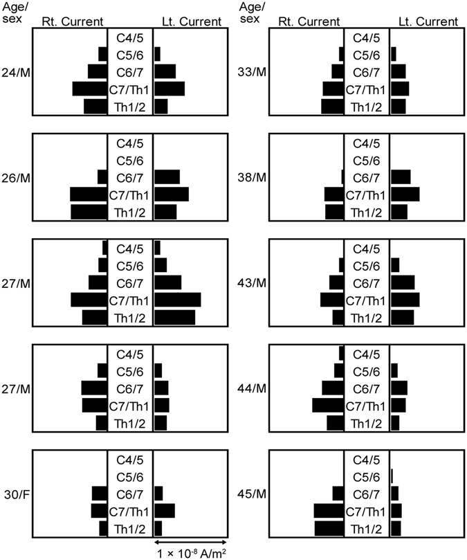
Diagnosis of nervous system disease is greatly aided by functional assessments and imaging techniques that localize neural activity abnormalities. Electrophysiological methods are helpful but often insufficient to locate neural lesions precisely. One proposed noninvasive alternative is magnetoneurography (MNG); we have developed MNG of the spinal cord (magnetospinography, MSG). Using a 120-channel superconducting quantum interference device biomagnetometer system in a magnetically shielded room, cervical spinal cord evoked magnetic fields (SCEFs) were recorded after stimulation of the lower thoracic cord in healthy subjects and a patient with cervical spondylotic myelopathy and after median nerve stimulation in healthy subjects. Electrophysiological activities in the spinal cord were reconstructed from SCEFs and visualized by a spatial filter, a recursive null-steering beamformer. Here, we show for the first time that MSG with high spatial and temporal resolution can be used to map electrophysiological activities in the cervical spinal cord and spinal nerve.
神经系统疾病的诊断极大地受益于功能评估和成像技术,这些技术可以定位神经活动异常。电生理学方法很有帮助,但往往不足以精确定位神经病变。一种被提议的非侵入性替代方法是磁神经图(MNG);我们已经开发了脊髓的 MNG(磁自旋图,MSG)。在磁屏蔽室中使用 120 通道超导量子干涉器件生物磁强计系统,在健康受试者和患有颈椎病性脊髓病的患者的下胸段脊髓刺激后以及在健康受试者的正中神经刺激后记录颈脊髓诱发磁场(SCEF)。通过空间滤波器(递归零导向波束形成器)从 SCEF 重建脊髓中的电生理活动并可视化。在这里,我们首次表明,具有高空间和时间分辨率的 MSG 可用于绘制颈脊髓和脊神经中的电生理活动。