Division of Neuroscience and Experimental Psychology, School of Biology, Faculty of Biology, Medicine and Health, University of Manchester, Manchester M13 9PL, UK.
Division of Neuroscience and Experimental Psychology, School of Biology, Faculty of Biology, Medicine and Health, University of Manchester, Manchester M13 9PL, UK.
Curr Biol. 2017 Jun 5;27(11):1623-1632.e4. doi: 10.1016/j.cub.2017.04.046. Epub 2017 May 18.
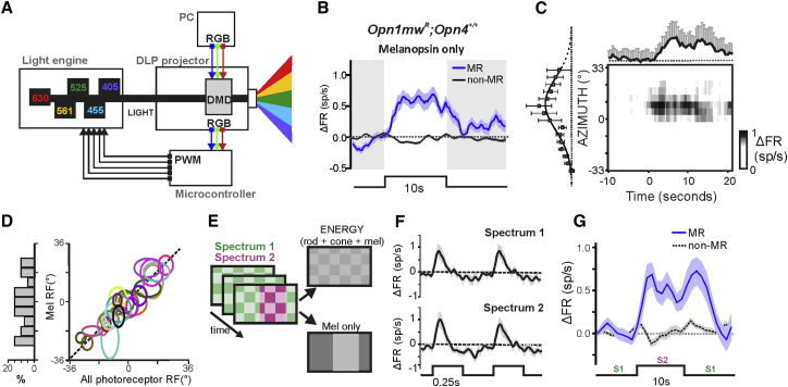
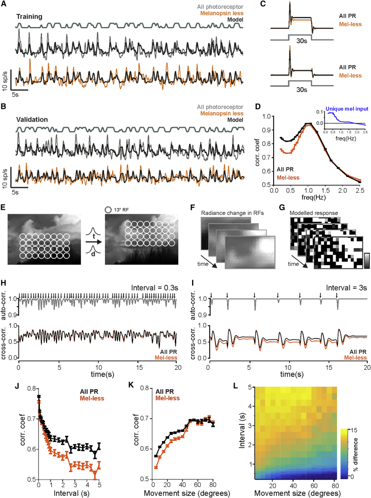
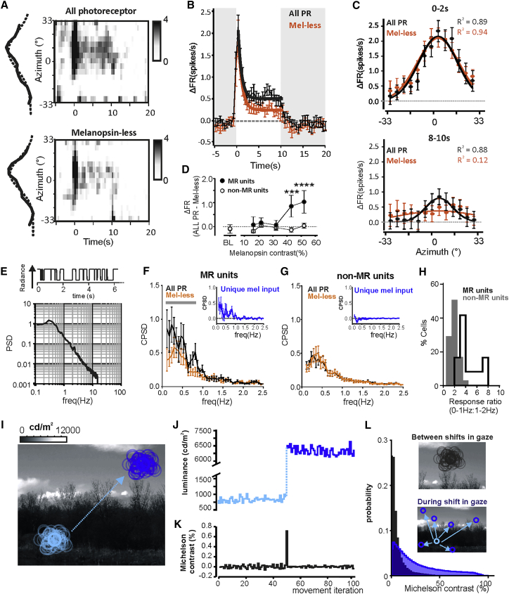
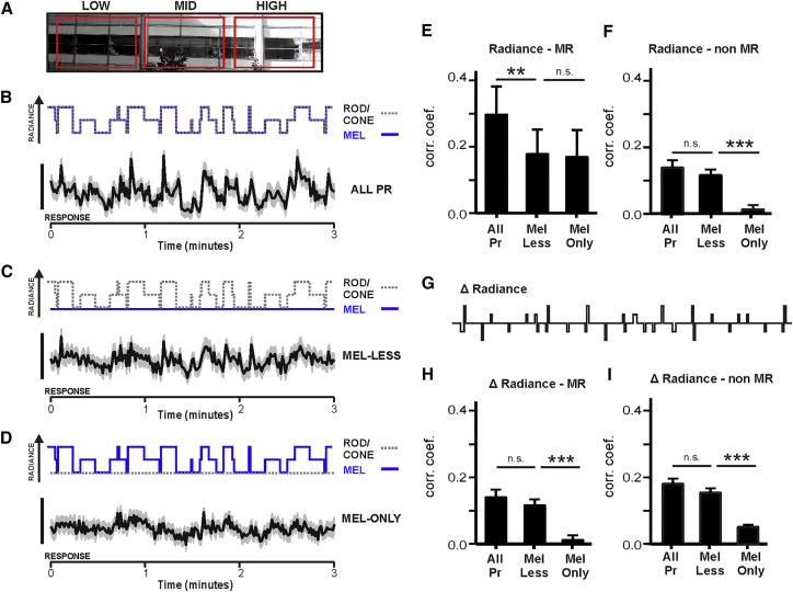
Melanopsin photoreception enhances retinal responses to variations in ambient light (irradiance) and drives non-image-forming visual reflexes such as circadian entrainment [1-6]. Melanopsin signals also reach brain regions responsible for form vision [7-9], but melanopsin's contribution, if any, to encoding visual images remains unclear. We addressed this deficit using principles of receptor silent substitution to present images in which visibility for melanopsin versus rods+cones was independently modulated, and we recorded evoked responses in the mouse dorsal lateral geniculate nucleus (dLGN; thalamic relay for cortical vision). Approximately 20% of dLGN units responded to patterns visible only to melanopsin, revealing that melanopsin signals alone can convey spatial information. Spatial receptive fields (RFs) mapped using melanopsin-isolating stimuli had ON centers with diameters ∼13°. Melanopsin and rod+cone responses differed in the temporal domain, and responses to slow changes in radiance (<0.9 Hz) and stationary images were deficient when stimuli were rendered invisible for melanopsin. We employed these data to devise and test a mathematical model of melanopsin's involvement in form vision and applied it, along with further experimental recordings, to explore melanopsin signals under simulated active view of natural scenes. Our findings reveal that melanopsin enhances the thalamic representation of scenes containing local correlations in radiance, compensating for the high temporal frequency bias of cone vision and the negative correlation between magnitude and frequency for changes in direction of view. Together, these data reveal a distinct melanopsin contribution to encoding visual images, predicting that, under natural view, melanopsin augments the early visual system's ability to encode patterns over moderate spatial scales.
褪黑素感光作用增强了视网膜对环境光(辐照度)变化的反应,并驱动非成像视觉反射,如昼夜节律的同步[1-6]。褪黑素信号也到达负责形态视觉的大脑区域[7-9],但褪黑素对编码视觉图像的贡献(如果有的话)仍然不清楚。我们使用受体沉默替代的原理来解决这一缺陷,即在独立调节褪黑素与视杆+视锥可见性的情况下呈现图像,我们记录了小鼠背外侧膝状体(dLGN;皮质视觉的丘脑中继)中的诱发反应。大约 20%的 dLGN 单位对仅能被褪黑素看到的模式有反应,这表明褪黑素信号本身可以传递空间信息。使用褪黑素隔离刺激映射的空间感受野(RFs)具有直径约为 13°的 ON 中心。褪黑素和视杆+视锥的反应在时域上有所不同,当刺激对褪黑素不可见时,对亮度缓慢变化(<0.9 Hz)和静止图像的反应就会不足。我们利用这些数据设计并测试了一个关于褪黑素参与形态视觉的数学模型,并应用它,以及进一步的实验记录,来探索模拟自然场景主动观察下的褪黑素信号。我们的发现表明,褪黑素增强了包含亮度局部相关性的场景在丘脑的表示,补偿了视锥视觉的高时间频率偏差,以及视角变化的幅度和频率之间的负相关。总的来说,这些数据揭示了褪黑素对编码视觉图像的独特贡献,预测在自然观察下,褪黑素增强了早期视觉系统在中等空间尺度上编码模式的能力。