Biophotonics Laboratory, Institute of Atomic Physics and Spectroscopy, University of Latvia, Riga LV-1586, Latvia.
Sensors (Basel). 2017 May 19;17(5):1165. doi: 10.3390/s17051165.
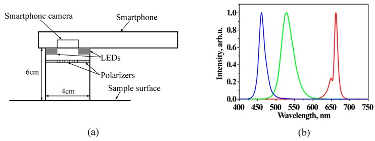
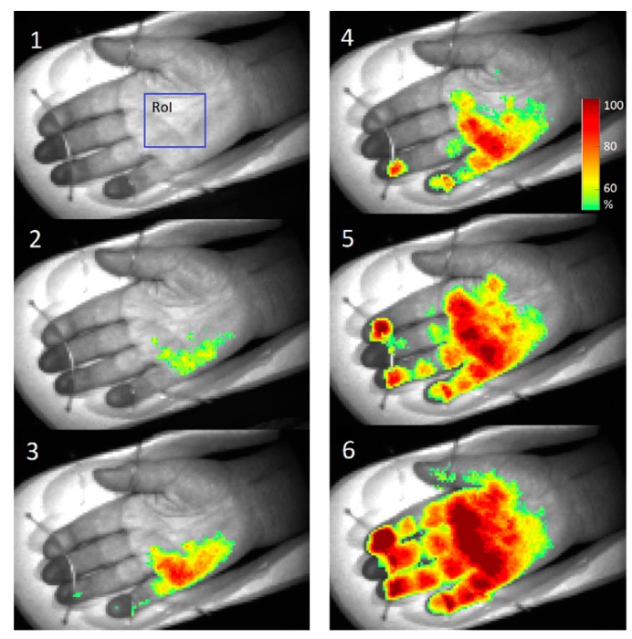
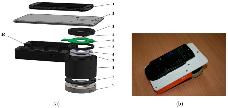
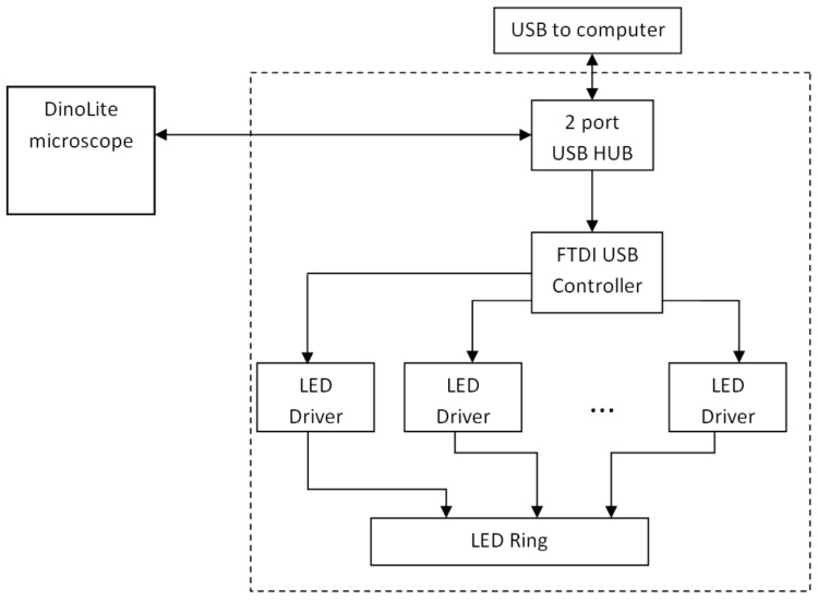
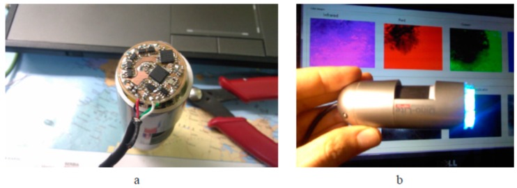
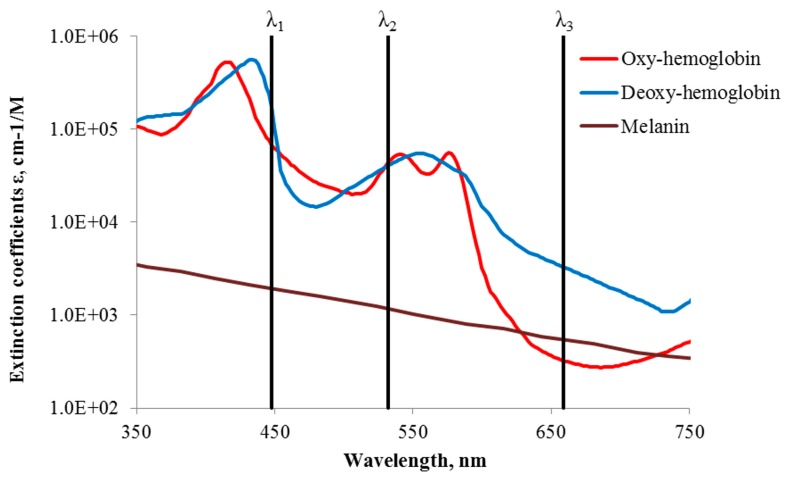
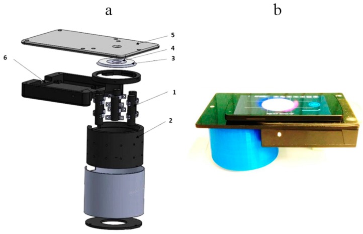
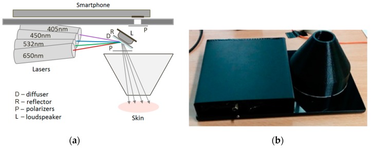
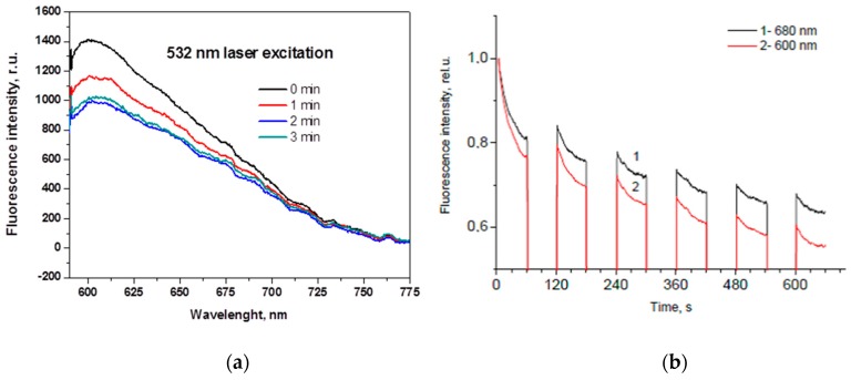
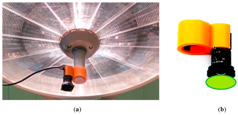
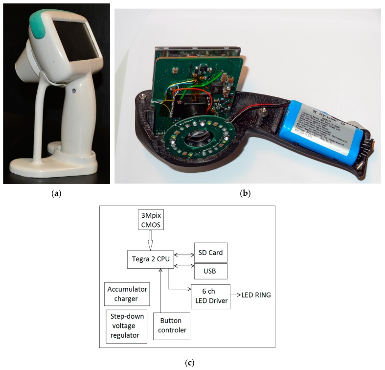
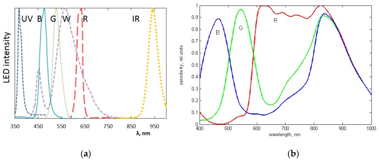
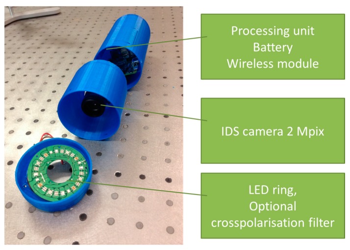
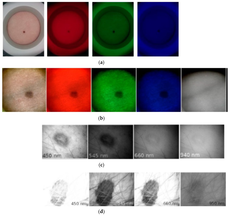
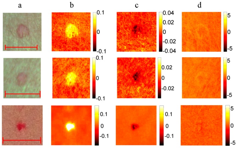
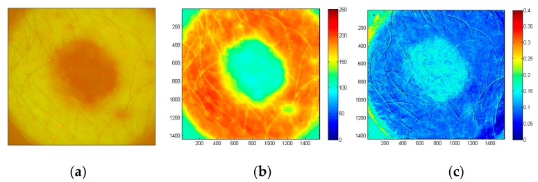
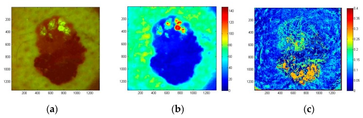
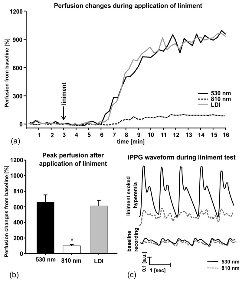
Optical tissue imaging has several advantages over the routine clinical imaging methods, including non-invasiveness (it does not change the structure of tissues), remote operation (it avoids infections) and the ability to quantify the tissue condition by means of specific image parameters. Dermatologists and other skin experts need compact (preferably pocket-size), self-sustaining and easy-to-use imaging devices. The operational principles and designs of ten portable in-vivo skin imaging prototypes developed at the Biophotonics Laboratory of Institute of Atomic Physics and Spectroscopy, University of Latvia during the recent five years are presented in this paper. Four groups of imaging devices are considered. Multi-spectral imagers offer possibilities for distant mapping of specific skin parameters, thus facilitating better diagnostics of skin malformations. Autofluorescence intensity and photobleaching rate imagers show a promising potential for skin tumor identification and margin delineation. Photoplethysmography video-imagers ensure remote detection of cutaneous blood pulsations and can provide real-time information on cardiovascular parameters and anesthesia efficiency. Multimodal skin imagers perform several of the abovementioned functions by taking a number of spectral and video images with the same image sensor. Design details of the developed prototypes and results of clinical tests illustrating their functionality are presented and discussed.
光学组织成像相对于常规临床成像方法具有多个优势,包括非侵入性(不会改变组织结构)、远程操作(避免感染)以及通过特定图像参数量化组织状况的能力。皮肤科医生和其他皮肤专家需要紧凑(最好是口袋大小)、自给自足且易于使用的成像设备。本文介绍了拉脱维亚大学原子物理与光谱学研究所生物光子学实验室在过去五年中开发的十种便携式体内皮肤成像原型的工作原理和设计。考虑了四组成像设备。多光谱成像仪提供了特定皮肤参数的远程映射可能性,从而有助于更好地诊断皮肤畸形。自发荧光强度和光漂白率成像仪显示出用于皮肤肿瘤识别和边界描绘的有希望的潜力。光体积描记术视频成像仪可确保远程检测皮肤搏动,并可提供有关心血管参数和麻醉效率的实时信息。多模态皮肤成像仪通过使用同一图像传感器拍摄多个光谱和视频图像来执行上述多项功能。本文介绍并讨论了所开发原型的设计细节以及临床测试结果,这些结果说明了其功能。