Pellegrini Erika, Signor Luca, Singh Saurabh, Boeri Erba Elisabetta, Cusack Stephen
European Molecular Biology Laboratory, Grenoble, France.
University Grenoble Alpes, IBS, Grenoble, France.
PLoS One. 2017 May 18;12(5):e0177161. doi: 10.1371/journal.pone.0177161. eCollection 2017.
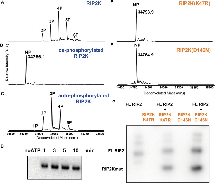
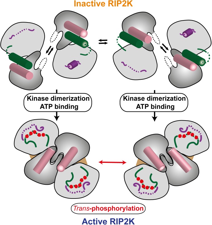
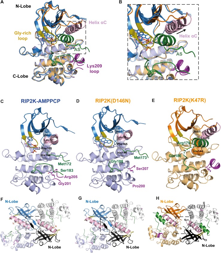
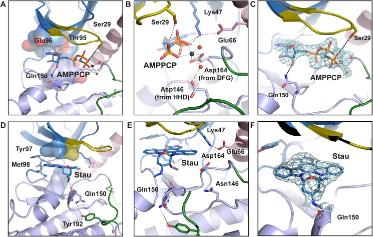
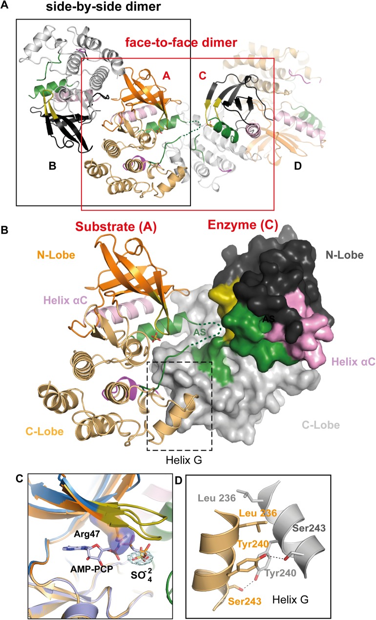
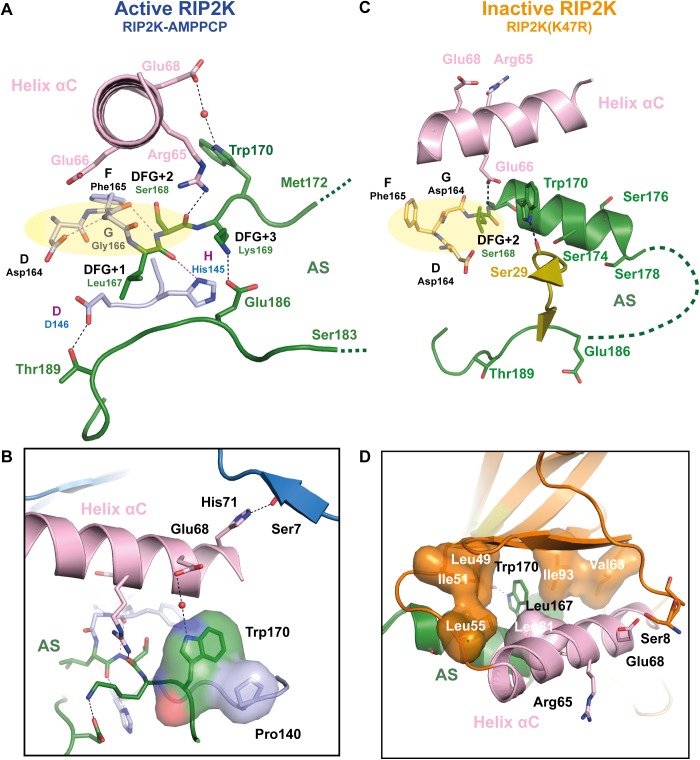
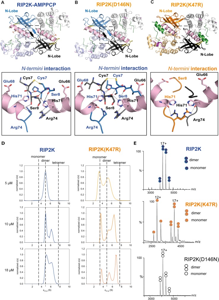
Innate immune receptors NOD1 and NOD2 are activated by bacterial peptidoglycans leading to recruitment of adaptor kinase RIP2, which, upon phosphorylation and ubiquitination, becomes a scaffold for downstream effectors. The kinase domain (RIP2K) is a pharmaceutical target for inflammatory diseases caused by aberrant NOD2-RIP2 signalling. Although structures of active RIP2K in complex with inhibitors have been reported, the mechanism of RIP2K activation remains to be elucidated. Here we analyse RIP2K activation by combining crystal structures of the active and inactive states with mass spectrometric characterization of their phosphorylation profiles. The active state has Helix αC inwardly displaced and the phosphorylated Activation Segment (AS) disordered, whilst in the inactive state Helix αC is outwardly displaced and packed against the helical, non-phosphorylated AS. Biophysical measurements show that the active state is a stable dimer whilst the inactive kinase is in a monomer-dimer equilibrium, consistent with the observed structural differences at the dimer interface. We conclude that RIP2 kinase auto-phosphorylation is intimately coupled to dimerization, similar to the case of BRAF. Our results will help drug design efforts targeting RIP2 as a potential treatment for NOD2-RIP2 related inflammatory diseases.
天然免疫受体NOD1和NOD2被细菌肽聚糖激活,导致衔接子激酶RIP2的募集,RIP2在磷酸化和泛素化后成为下游效应器的支架。激酶结构域(RIP2K)是由异常的NOD2-RIP2信号传导引起的炎症性疾病的药物靶点。尽管已经报道了活性RIP2K与抑制剂复合物的结构,但其激活机制仍有待阐明。在这里,我们通过结合活性和非活性状态的晶体结构及其磷酸化谱的质谱表征来分析RIP2K的激活。活性状态下,螺旋αC向内位移,磷酸化的激活片段(AS)无序,而在非活性状态下,螺旋αC向外位移并与螺旋状的、未磷酸化的AS堆积在一起。生物物理测量表明,活性状态是稳定的二聚体,而非活性激酶处于单体-二聚体平衡,这与在二聚体界面观察到的结构差异一致。我们得出结论,RIP2激酶的自磷酸化与二聚化密切相关,类似于BRAF的情况。我们的结果将有助于针对RIP2的药物设计工作,作为治疗NOD2-RIP2相关炎症性疾病的潜在方法。