Sakamoto Kazuo, Nozoe Masatsugu, Tsutsui Yoshitomo, Suematsu Nobuhiro, Kubota Toru, Okabe Masanori, Yamamoto Yusuke
Division of Cardiology, Cardiovascular and Aortic Center, Saiseikai Fukuoka General Hospital, Fukuoka, Japan.
Int Med Case Rep J. 2017 May 11;10:167-171. doi: 10.2147/IMCRJ.S135952. eCollection 2017.
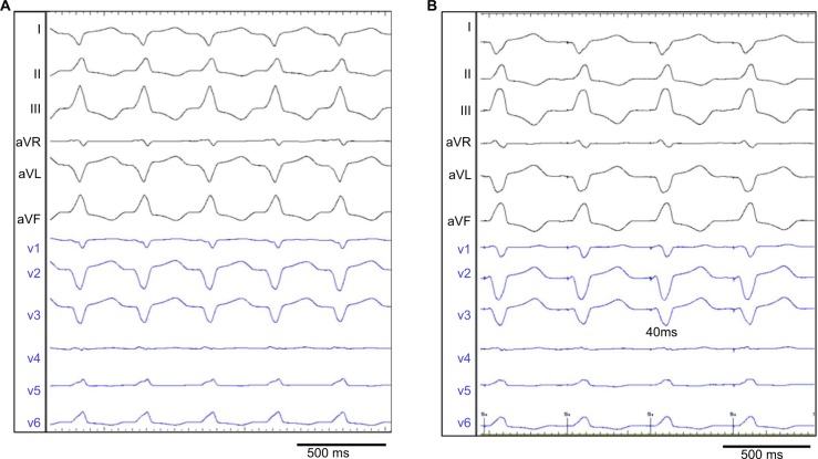
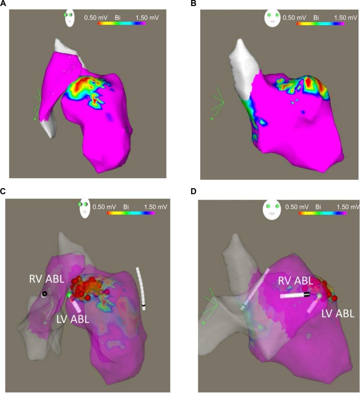
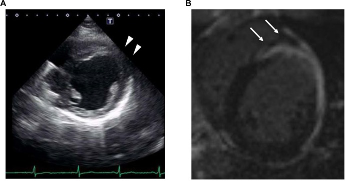
Cardiac magnetic resonance imaging (MRI) is a useful tool for detecting the arrhythmogenic substrate in cardiac sarcoidosis. We herein present a case of bipolar radiofrequency catheter ablation for ventricular tachycardia (VT) complicated with cardiac sarcoidosis, guided by pre-procedural cardiac MRI. Neither echocardiography nor endocardial voltage mapping suggested a septal VT substrate. However, MRI alone detected intramural lesions in the septum. Although application of endocardial energy failed to treat the VT, bipolar ablation targeting the potential substrate identified by MRI successfully eliminated the VT. Even when no abnormalities are depicted on echocardiography and endocardial voltage mapping, intramural scar tissue identified by cardiac MRI could be critical for VT.
心脏磁共振成像(MRI)是检测心脏结节病中致心律失常基质的有用工具。我们在此介绍一例在术前心脏MRI引导下,对合并心脏结节病的室性心动过速(VT)进行双极射频导管消融的病例。超声心动图和心内膜电压标测均未提示室间隔VT基质。然而,仅MRI检测到室间隔壁内病变。尽管心内膜能量应用未能治疗VT,但针对MRI确定的潜在基质进行双极消融成功消除了VT。即使超声心动图和心内膜电压标测未显示异常,心脏MRI确定的壁内瘢痕组织对VT可能也至关重要。