• 文献检索
  • 文档翻译
  • 深度研究
  • 学术资讯
  • Suppr Zotero 插件Zotero 插件
  • 邀请有礼
  • 套餐&价格
  • 历史记录
应用&插件
Suppr Zotero 插件Zotero 插件浏览器插件Mac 客户端Windows 客户端微信小程序
定价
高级版会员购买积分包购买API积分包
服务
文献检索文档翻译深度研究API 文档MCP 服务
关于我们
关于 Suppr公司介绍联系我们用户协议隐私条款
关注我们

Suppr 超能文献

核心技术专利:CN118964589B侵权必究
粤ICP备2023148730 号-1Suppr @ 2026

文献检索

告别复杂PubMed语法,用中文像聊天一样搜索,搜遍4000万医学文献。AI智能推荐,让科研检索更轻松。

立即免费搜索

文件翻译

保留排版,准确专业,支持PDF/Word/PPT等文件格式,支持 12+语言互译。

免费翻译文档

深度研究

AI帮你快速写综述,25分钟生成高质量综述,智能提取关键信息,辅助科研写作。

立即免费体验

应激心脏磁共振灌注成像前摄入咖啡因对通过T1映射测量的瑞加腺苷与腺苷诱导的充血的影响。

Effects of caffeine intake prior to stress cardiac magnetic resonance perfusion imaging on regadenoson- versus adenosine-induced hyperemia as measured by T1 mapping.

作者信息

van Dijk R, Kuijpers D, Kaandorp T A M, van Dijkman P R M, Vliegenthart R, van der Harst P, Oudkerk M

机构信息

Center for Medical Imaging, University Medical Center Groningen, University of Groningen, Hanzeplein 1 EB 45, Groningen, The Netherlands.

Department of Cardiology, University Medical Center Groningen, University of Groningen, Groningen, The Netherlands.

出版信息

Int J Cardiovasc Imaging. 2017 Nov;33(11):1753-1759. doi: 10.1007/s10554-017-1157-4. Epub 2017 May 25.

DOI:10.1007/s10554-017-1157-4
PMID:28547666
原文链接:https://pmc.ncbi.nlm.nih.gov/articles/PMC5682854/
Abstract

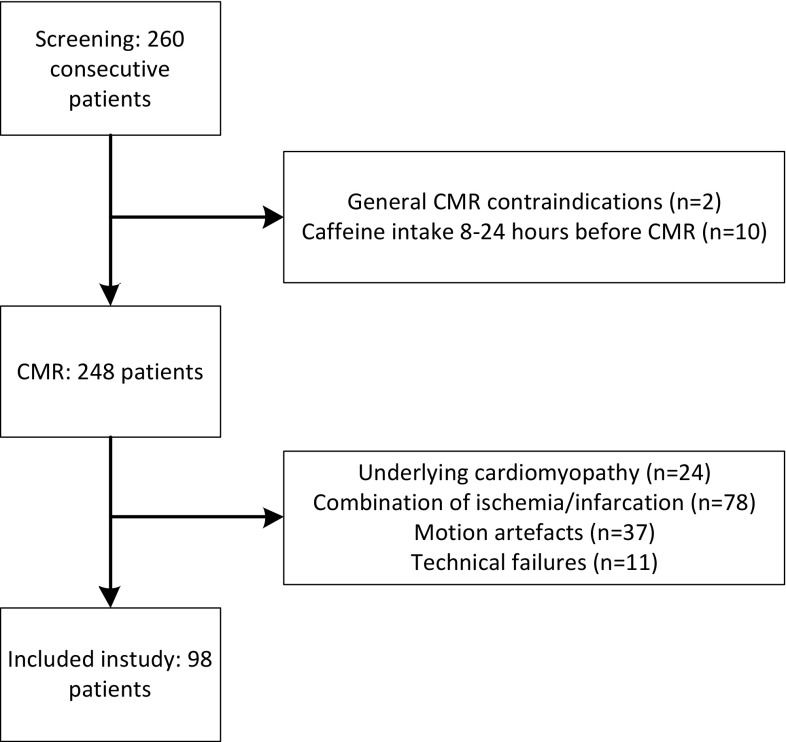
The antagonistic effects of caffeine on adenosine receptors are a possible cause of false-negative stress perfusion imaging. The purpose of this study was to determine the effects of coffee intake <4 h prior to stress perfusion cardiac magnetic resonance imaging (CMR) in regadenoson- versus adenosine-induced hyperemia as measured with T1-mapping. 98 consecutive patients with suspected coronary artery disease referred for either adenosine or regadenoson perfusion CMR were included in this analysis. Twenty-four patients reported coffee consumption <4 h before CMR (15 patients with adenosine, and 9 patients with regadenoson); 74 patients reported no coffee intake (50 patients with adenosine, and 24 patients with regadenoson). T1 mapping was performed using a modified look-locker inversion recovery sequence. T1 reactivity was determined by subtracting T1 from T1. T1, T1, and T1 reactivity in patients referred for regadenoson perfusion CMR were not significantly different when comparing patients with <4 h coffee intake and patients who reported no coffee intake (976 ± 4 ms, 1019 ± 48 ms, and 4.4 ± 3.2% vs 971 ± 33 ms, 1023 ± 43 ms, and 5.4 ± 2.4%) (p = 0.70, 0.79, and 0.40), and similar to values in patients without coffee intake undergoing adenosine CMR. In patients with <4 h coffee intake, T1, and T1 reactivity were significantly lower for adenosine (898 ± 51 ms, and -7.8 ± 5.0%) compared to regadenoson perfusion CMR (p < 0.001). Coffee intake <4 h prior to regadenoson perfusion CMR has no effect on stress-induced hyperemia as measured with T1 mapping.

摘要

咖啡因对腺苷受体的拮抗作用可能是导致应激灌注成像假阴性的原因。本研究的目的是确定在使用T1映射测量瑞加腺苷和腺苷诱发的充血时,在应激灌注心脏磁共振成像(CMR)前<4小时摄入咖啡的影响。本分析纳入了98例因腺苷或瑞加腺苷灌注CMR而被转诊的疑似冠心病患者。24例患者报告在CMR前<4小时饮用了咖啡(15例接受腺苷检查,9例接受瑞加腺苷检查);74例患者报告未摄入咖啡(50例接受腺苷检查,24例接受瑞加腺苷检查)。使用改良的锁相环反转恢复序列进行T1映射。T1反应性通过从T1中减去T1来确定。在比较<4小时咖啡摄入量的患者和未摄入咖啡的患者时,接受瑞加腺苷灌注CMR的患者的T1、T1和T1反应性无显著差异(分别为976±4毫秒、1019±48毫秒和4.4±3.2%,对比971±33毫秒、1023±43毫秒和5.4±2.4%)(p = 0.70、0.79和0.40),且与未摄入咖啡而接受腺苷CMR检查的患者的值相似。在<4小时咖啡摄入量的患者中,与瑞加腺苷灌注CMR相比,腺苷的T1和T1反应性显著更低(分别为898±51毫秒和-7.8±5.0%)(p < 0.001)。在瑞加腺苷灌注CMR前<4小时摄入咖啡对用T1映射测量的应激诱发充血无影响。

https://cdn.ncbi.nlm.nih.gov/pmc/blobs/926b/5682854/8da4ea105895/10554_2017_1157_Fig3_HTML.jpg
https://cdn.ncbi.nlm.nih.gov/pmc/blobs/926b/5682854/760180adb9eb/10554_2017_1157_Fig1_HTML.jpg
https://cdn.ncbi.nlm.nih.gov/pmc/blobs/926b/5682854/0370294d5e0e/10554_2017_1157_Fig2_HTML.jpg
https://cdn.ncbi.nlm.nih.gov/pmc/blobs/926b/5682854/8da4ea105895/10554_2017_1157_Fig3_HTML.jpg
https://cdn.ncbi.nlm.nih.gov/pmc/blobs/926b/5682854/760180adb9eb/10554_2017_1157_Fig1_HTML.jpg
https://cdn.ncbi.nlm.nih.gov/pmc/blobs/926b/5682854/0370294d5e0e/10554_2017_1157_Fig2_HTML.jpg
https://cdn.ncbi.nlm.nih.gov/pmc/blobs/926b/5682854/8da4ea105895/10554_2017_1157_Fig3_HTML.jpg

相似文献

1
Effects of caffeine intake prior to stress cardiac magnetic resonance perfusion imaging on regadenoson- versus adenosine-induced hyperemia as measured by T1 mapping.应激心脏磁共振灌注成像前摄入咖啡因对通过T1映射测量的瑞加腺苷与腺苷诱导的充血的影响。
Int J Cardiovasc Imaging. 2017 Nov;33(11):1753-1759. doi: 10.1007/s10554-017-1157-4. Epub 2017 May 25.
2
Caffeine intake inverts the effect of adenosine on myocardial perfusion during stress as measured by T1 mapping.通过T1映射测量发现,在应激状态下,咖啡因摄入会逆转腺苷对心肌灌注的影响。
Int J Cardiovasc Imaging. 2016 Oct;32(10):1545-53. doi: 10.1007/s10554-016-0949-2. Epub 2016 Jul 29.
3
Disagreement between splenic switch-off and myocardial T1-mapping after caffeine intake.摄入咖啡因后脾脏关闭与心肌T1映射之间的差异。
Int J Cardiovasc Imaging. 2018 Apr;34(4):625-632. doi: 10.1007/s10554-017-1274-0. Epub 2017 Nov 24.
4
Impact of caffeine on myocardial perfusion reserve assessed by semiquantitative adenosine stress perfusion cardiovascular magnetic resonance.腺苷负荷半定量心肌灌注磁共振评估咖啡因对心肌灌注储备的影响。
J Cardiovasc Magn Reson. 2019 Jun 24;21(1):33. doi: 10.1186/s12968-019-0542-7.
5
Rapid rest/stress regadenoson ungated perfusion CMR for detection of coronary artery disease in patients with atrial fibrillation.用于检测心房颤动患者冠状动脉疾病的快速静息/负荷瑞加诺生非门控灌注心脏磁共振成像
Int J Cardiovasc Imaging. 2017 Nov;33(11):1781-1788. doi: 10.1007/s10554-017-1168-1. Epub 2017 May 20.
6
Considerations when measuring myocardial perfusion reserve by cardiovascular magnetic resonance using regadenoson.考虑使用雷卡地诺生测量心血管磁共振心肌灌注储备时的注意事项。
J Cardiovasc Magn Reson. 2012 Dec 28;14(1):89. doi: 10.1186/1532-429X-14-89.
7
The effect of obesity on regadenoson-induced myocardial hyperemia: a quantitative magnetic resonance imaging study.肥胖对瑞加德松诱导的心肌充血的影响:一项定量磁共振成像研究。
Int J Cardiovasc Imaging. 2012 Aug;28(6):1435-44. doi: 10.1007/s10554-011-9949-4. Epub 2011 Oct 4.
8
Prognostic value of normal regadenoson stress perfusion cardiovascular magnetic resonance.正常 regadenoson 应激灌注心血管磁共振的预后价值。
J Cardiovasc Magn Reson. 2013 Dec 21;15(1):108. doi: 10.1186/1532-429X-15-108.
9
Effects of Caffeine on Myocardial Blood Flow: A Systematic Review.咖啡因对心肌血流的影响:系统评价。
Nutrients. 2018 Aug 13;10(8):1083. doi: 10.3390/nu10081083.
10
Adenosine stress CMR T1-mapping detects early microvascular dysfunction in patients with type 2 diabetes mellitus without obstructive coronary artery disease.腺苷负荷心脏磁共振 T1 mapping 检测 2 型糖尿病无阻塞性冠状动脉疾病患者的早期微血管功能障碍。
J Cardiovasc Magn Reson. 2017 Oct 25;19(1):81. doi: 10.1186/s12968-017-0397-8.

引用本文的文献

1
Safety, feasibility, and hemodynamic response of regadenoson for stress perfusion CMR.瑞加德松用于应激灌注 CMR的安全性、可行性和血液动力学反应。
Int J Cardiovasc Imaging. 2023 Sep;39(9):1765-1774. doi: 10.1007/s10554-023-02877-z. Epub 2023 Jun 24.
2
Cardiovascular Magnetic Resonance Parametric Mapping Techniques for the Assessment of Chronic Coronary Syndromes.用于评估慢性冠状动脉综合征的心血管磁共振参数成像技术
J Cardiovasc Dev Dis. 2022 Dec 9;9(12):443. doi: 10.3390/jcdd9120443.
3
Quantitative myocardial perfusion response to adenosine and regadenoson in patients with suspected coronary artery disease.

本文引用的文献

1
Low, but not high, dose caffeine is a readily available probe for adenosine actions.低剂量而非高剂量的咖啡因是一种易于获得的腺苷作用探针。
Mol Aspects Med. 2017 Jun;55:20-25. doi: 10.1016/j.mam.2016.11.011. Epub 2016 Nov 30.
2
Caffeine intake inverts the effect of adenosine on myocardial perfusion during stress as measured by T1 mapping.通过T1映射测量发现,在应激状态下,咖啡因摄入会逆转腺苷对心肌灌注的影响。
Int J Cardiovasc Imaging. 2016 Oct;32(10):1545-53. doi: 10.1007/s10554-016-0949-2. Epub 2016 Jul 29.
3
Caffeine does not significantly reduce the sensitivity of vasodilator stress MPI: Rebuttal.
腺苷和雷卡地诺生诱导的心肌灌注定量反应在疑似冠心病患者中的变化。
J Nucl Cardiol. 2022 Feb;29(1):24-36. doi: 10.1007/s12350-021-02731-6. Epub 2021 Aug 12.
4
Cardiac stress T1-mapping response and extracellular volume stability of MOLLI-based T1-mapping methods.基于 MOLLI 的 T1 映射方法的心脏应激 T1 映射反应和细胞外容积稳定性。
Sci Rep. 2021 Jun 30;11(1):13568. doi: 10.1038/s41598-021-92923-4.
5
Significant Impact of Coffee Consumption on MR-Based Measures of Cardiac Function in a Population-Based Cohort Study without Manifest Cardiovascular Disease.在一项无明显心血管疾病的人群队列研究中,咖啡消费对基于磁共振成像的心脏功能测量指标有显著影响。
Nutrients. 2021 Apr 13;13(4):1275. doi: 10.3390/nu13041275.
6
Molecular Mechanisms of Adenosine Stress T1 Mapping.腺苷应激 T1 映射的分子机制。
Circ Cardiovasc Imaging. 2021 Mar;14(3):e011774. doi: 10.1161/CIRCIMAGING.120.011774. Epub 2021 Mar 12.
7
Regadenoson Stress Testing: A Comprehensive Review With a Focused Update.雷加得诺素负荷试验:一项重点更新的综合综述
Cureus. 2021 Jan 27;13(1):e12940. doi: 10.7759/cureus.12940.
8
Feature tracking and mapping analysis of myocardial response to improved perfusion reserve in patients with refractory angina treated by coronary sinus Reducer implantation: a CMR study.经冠状窦内逆行减容术治疗难治性心绞痛患者心肌对改善灌注储备反应的特征跟踪和绘图分析:一项 CMR 研究。
Int J Cardiovasc Imaging. 2021 Jan;37(1):291-303. doi: 10.1007/s10554-020-01964-9. Epub 2020 Aug 28.
9
Computed tomography for myocardial characterization in ischemic heart disease: a state-of-the-art review.计算机断层扫描心肌特征化在缺血性心脏病中的应用:最新综述。
Eur Radiol Exp. 2020 Jun 17;4(1):36. doi: 10.1186/s41747-020-00158-1.
10
The reliability and feasibility of non-contrast adenosine stress cardiovascular magnetic resonance T1 mapping in patients on haemodialysis.非对比剂腺苷负荷心血管磁共振T1成像在血液透析患者中的可靠性和可行性
J Cardiovasc Magn Reson. 2020 Jun 8;22(1):43. doi: 10.1186/s12968-020-00634-y.
咖啡因不会显著降低血管扩张应激心肌灌注成像的敏感性:反驳。
J Nucl Cardiol. 2016 Jun;23(3):604. doi: 10.1007/s12350-016-0421-7. Epub 2016 Feb 10.
4
Adenosine Stress and Rest T1 Mapping Can Differentiate Between Ischemic, Infarcted, Remote, and Normal Myocardium Without the Need for Gadolinium Contrast Agents.腺苷负荷与静息状态下的T1映射无需钆对比剂即可区分缺血、梗死、远隔和正常心肌。
JACC Cardiovasc Imaging. 2016 Jan;9(1):27-36. doi: 10.1016/j.jcmg.2015.08.018. Epub 2015 Dec 9.
5
Factors associated with false-negative cardiovascular magnetic resonance perfusion studies: A Clinical evaluation of magnetic resonance imaging in coronary artery disease (CE-MARC) substudy.心血管磁共振灌注研究假阴性相关因素:冠状动脉疾病磁共振成像临床评估(CE-MARC)子研究
J Magn Reson Imaging. 2016 Mar;43(3):566-73. doi: 10.1002/jmri.25032. Epub 2015 Aug 18.
6
Caffeine Consumption and Heart Rate and Blood Pressure Response to Regadenoson.咖啡因摄入与瑞加德松引起的心率及血压反应
PLoS One. 2015 Jun 22;10(6):e0130487. doi: 10.1371/journal.pone.0130487. eCollection 2015.
7
2014 ESC/EACTS guidelines on myocardial revascularization.2014年欧洲心脏病学会/欧洲心胸外科学会心肌血运重建指南。
Rev Esp Cardiol (Engl Ed). 2015 Feb;68(2):144. doi: 10.1016/j.rec.2014.12.006.
8
Effect of caffeine on SPECT myocardial perfusion imaging during regadenoson pharmacologic stress: a prospective, randomized, multicenter study.咖啡因对雷加得诺药理学应激期间单光子发射计算机断层扫描心肌灌注成像的影响:一项前瞻性、随机、多中心研究。
Int J Cardiovasc Imaging. 2014 Jun;30(5):979-89. doi: 10.1007/s10554-014-0419-7. Epub 2014 Apr 17.
9
Myocardial T1 mapping and extracellular volume quantification: a Society for Cardiovascular Magnetic Resonance (SCMR) and CMR Working Group of the European Society of Cardiology consensus statement.心肌 T1 映射和细胞外容积定量:心血管磁共振学会 (SCMR) 和欧洲心脏病学会 CMR 工作组的共识声明。
J Cardiovasc Magn Reson. 2013 Oct 14;15(1):92. doi: 10.1186/1532-429X-15-92.
10
Superior diagnostic performance of perfusion-cardiovascular magnetic resonance versus SPECT to detect coronary artery disease: The secondary endpoints of the multicenter multivendor MR-IMPACT II (Magnetic Resonance Imaging for Myocardial Perfusion Assessment in Coronary Artery Disease Trial).与单光子发射计算机断层扫描相比,灌注-心血管磁共振具有更好的诊断性能,可用于检测冠状动脉疾病:多中心多供应商磁共振影响 II 试验(磁共振成像在冠状动脉疾病中的心肌灌注评估试验)的次要终点。
J Cardiovasc Magn Reson. 2012 Sep 2;14(1):61. doi: 10.1186/1532-429X-14-61.