Krishnamurthy Pranathi Meda, Shukla Shirish, Ray Paramita, Mehra Rohit, Nyati Mukesh K, Lawrence Theodore S, Ray Dipankar
Department of Radiation Oncology, University of Michigan, Ann Arbor, MI, USA.
Current address: RNA Therapeutics Institute, University of Massachusetts Medical School, Worcester, MA, USA.
Oncotarget. 2017 Jul 18;8(29):47767-47779. doi: 10.18632/oncotarget.17770.
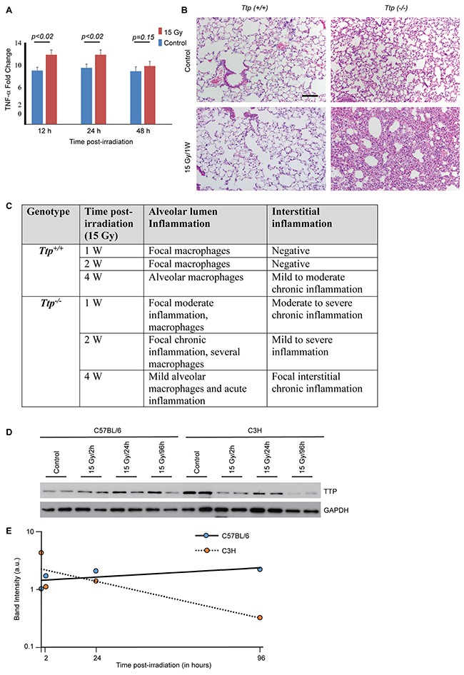
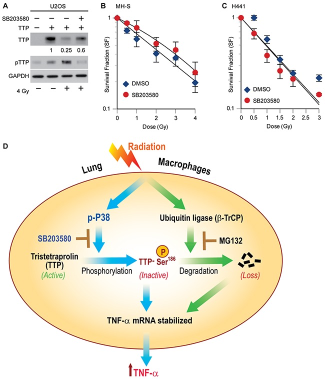
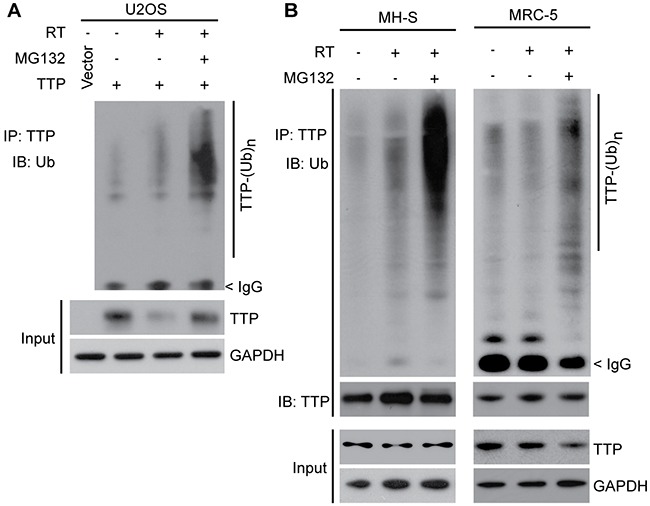
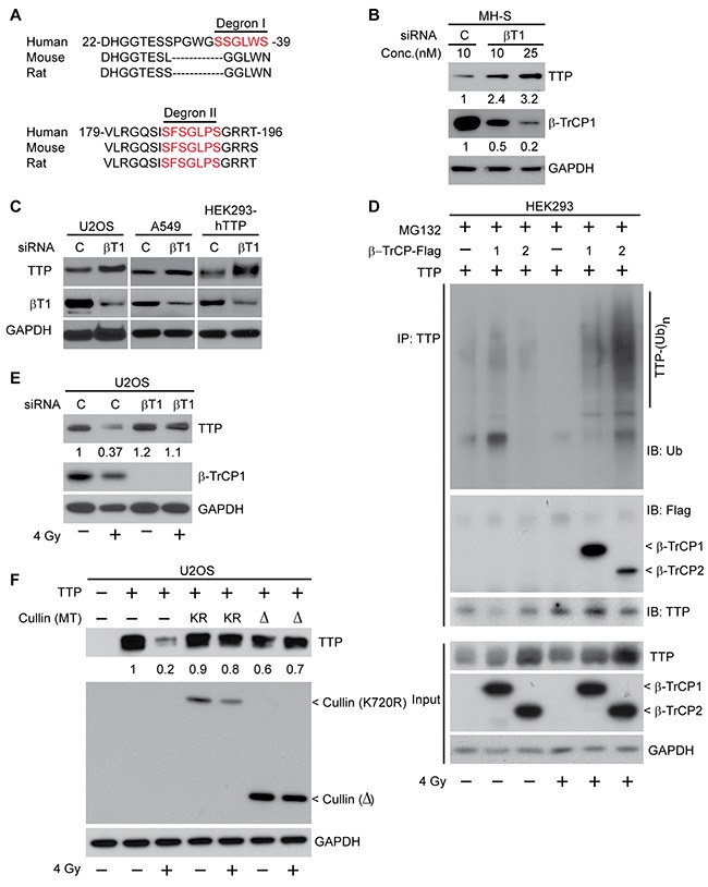
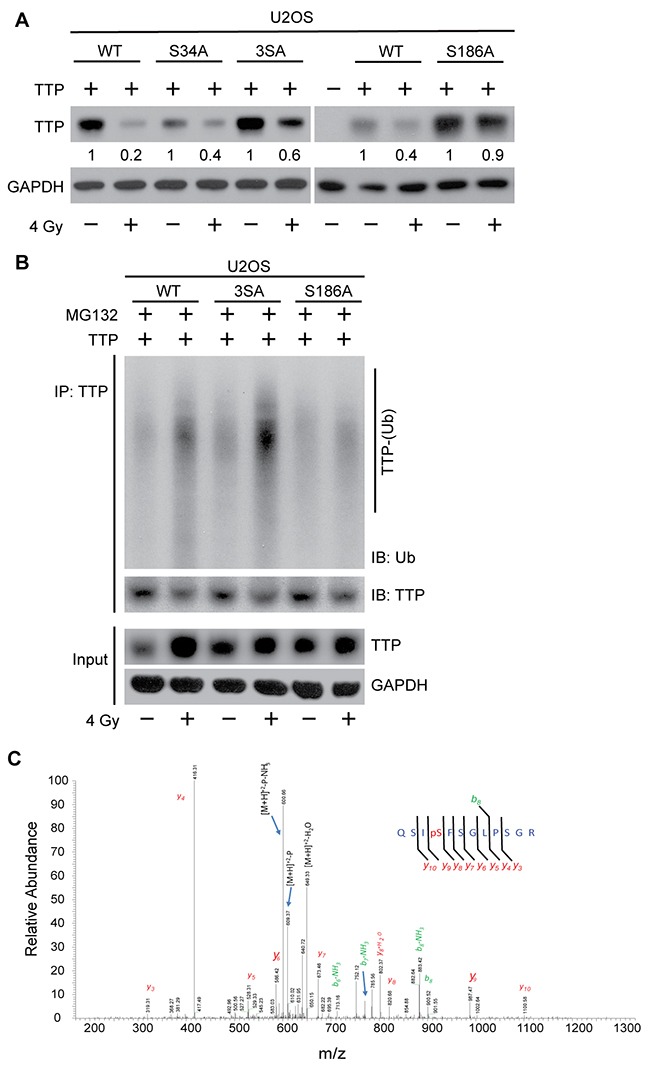
Early release of tumor necrosis factor-alpha (TNF-α) during radiotherapy of thoracic cancers plays an important role in radiation pneumonitis, whose inhibition may provide lung radioprotection. We previously reported radiation inactivates Tristetraprolin (TTP), a negative regulator of TNF-α synthesis, which correlated with increased TNF-α release. However, the molecular events involved in radiation-induced TTP inactivation remain unclear. To determine if eliminating Ttp in mice resulted in a phenotypic response to radiation, Ttp-null mice lungs were exposed to a single dose of 15 Gy, and TNF-α release and lung inflammation were analyzed at different time points post-irradiation. Ttp-/- mice with elevated (9.5±0.6 fold) basal TNF-α showed further increase (12.2±0.9 fold, p<0.02) in TNF-α release and acute lung inflammation within a week post-irradiation. Further studies using mouse lung macrophage (MH-S), human lung fibroblast (MRC-5), and exogenous human TTP overexpressing U2OS and HEK293 cells upon irradiation (a single dose of 4 Gy) promoted p38-mediated TTP phosphorylation at the serine 186 position, which primed it to be recognized by an ubiquitin ligase (E3), beta transducing repeat containing protein (β-TrCP), to promote polyubiquitination-mediated proteasomal degradation. Consequently, a serine 186 to alanine (SA) mutant of TTP was resistant to radiation-induced degradation. Similarly, either a p38 kinase inhibitor (SB203580), or siRNA-mediated β-TrCP knockdown, or overexpression of dominant negative Cullin1 mutants protected TTP from radiation-induced degradation. Consequently, SB203580 pretreatment blocked radiation-induced TNF-α release and radioprotected macrophages. Together, these data establish the involvement of the p38-βTrCP-TTP-TNFα signaling axis in radiation-induced lung inflammation and identified p38 inhibition as a possible lung radioprotection strategy.
在胸段癌放疗期间肿瘤坏死因子-α(TNF-α)的早期释放,在放射性肺炎中起重要作用,抑制该因子可能提供肺部放射保护作用。我们之前报道过,放疗会使TNF-α合成的负调节因子锌指蛋白36(TTP)失活,这与TNF-α释放增加相关。然而,放疗诱导TTP失活所涉及的分子事件仍不清楚。为了确定敲除小鼠体内的Ttp是否会导致对辐射的表型反应,将Ttp基因敲除小鼠的肺暴露于15 Gy的单剂量辐射下,并在照射后的不同时间点分析TNF-α释放和肺部炎症情况。基础TNF-α水平升高(9.5±0.6倍)的Ttp-/-小鼠,在照射后一周内TNF-α释放进一步增加(12.2±0.9倍,p<0.02),并出现急性肺部炎症。进一步的研究表明,对小鼠肺巨噬细胞(MH-S)、人肺成纤维细胞(MRC-5)以及照射(单剂量4 Gy)后过表达外源性人TTP的U-2 OS和HEK293细胞进行研究发现,p38会介导TTP在丝氨酸186位点发生磷酸化,使其能被泛素连接酶(E3)——含β转导重复序列蛋白(β-TrCP)识别,从而促进多聚泛素化介导的蛋白酶体降解。因此,TTP的丝氨酸186突变为丙氨酸(SA)的突变体对辐射诱导的降解具有抗性。同样,使用p38激酶抑制剂(SB203580)、siRNA介导的β-TrCP敲低或者显性负性Cullin1突变体的过表达,均可保护TTP免受辐射诱导的降解。因此,SB203580预处理可阻断辐射诱导的TNF-α释放,并对巨噬细胞起到放射保护作用。总之,这些数据证实了p38-βTrCP-TTP-TNFα信号轴参与了辐射诱导的肺部炎症,并确定抑制p38是一种可能的肺部放射保护策略。