Liu Jia, Zhang Bo, Li Maotong, Zhou Meijun, Li Fei, Huang Xiuxian, Pan Min, Xue Li, Yan Fei
Department of Echocardiography, Fourth Affiliated Hospital of Harbin Medical University, Harbin, Heilongjiang, China.
Department of Echocardiography, Shanghai Eastern Hospital Affiliated to Tongji University, Shanghai, China.
PLoS One. 2017 May 30;12(5):e0178031. doi: 10.1371/journal.pone.0178031. eCollection 2017.
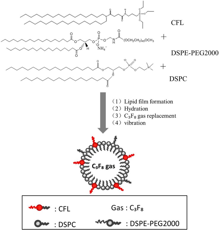
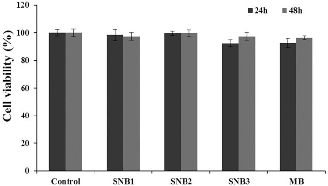
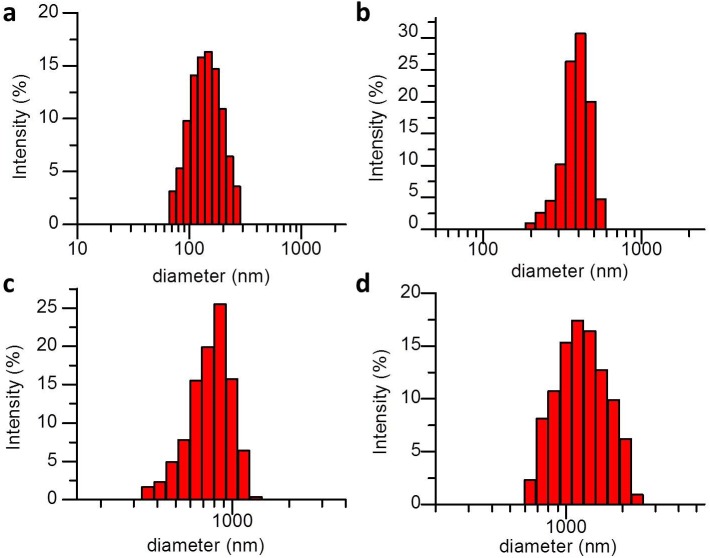
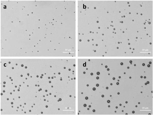
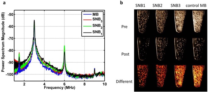
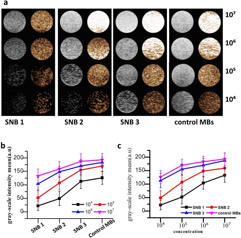
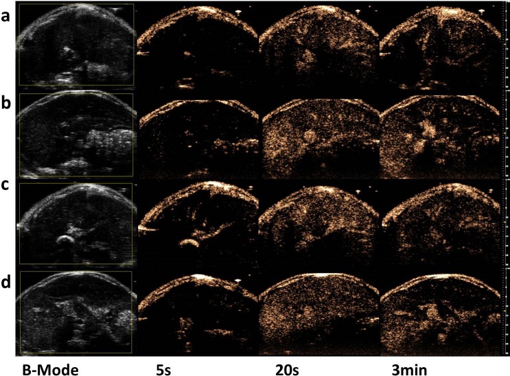
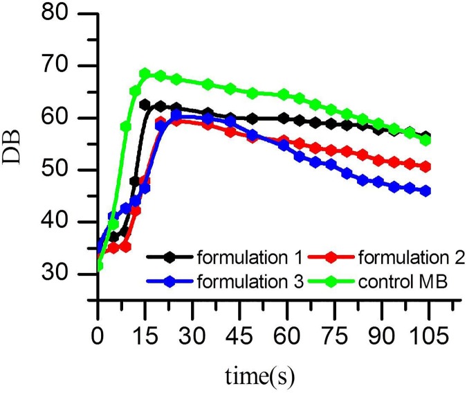
Nanobubbles (NBs) opened a new field of ultrasound imaging. There is still no practical method to control the diameter of bubbles. In this study, we developed a new method to control the size by incorporating of silicon hybrid lipids into the bubble membrane. The range of particle size of resulting NBs is between 523.02 ± 46.45 to 857.18 ± 82.90, smaller than the conventional microbubbles. The size of resulting NBs increased with the decrease in amount of silicon hybrid lipids, indicating the diameter of NBs can be regulated through modulating the ratio of silicon hybrid lipids in the bubble shell. Typical harmonic signals could be detected. The in vitro and in vivo ultrasound imaging experiments demonstrated these silicon-modified NBs had significantly improved ultrasound contrast enhancement abilities. Cytotoxicity assays revealed that these NBs had no obvious cytotoxicity to the 293 cell line at the tested bubble concentration. Our results showed that the novel NBs could use as nanoscale ultrasound contrast agents, providing the foundation for NBs in future applications including contrast-enhanced imaging and drug/gene delivery.
纳米气泡(NBs)开启了超声成像的新领域。目前仍没有控制气泡直径的实用方法。在本研究中,我们开发了一种通过将硅杂化脂质掺入气泡膜来控制尺寸的新方法。所得纳米气泡的粒径范围在523.02±46.45至857.18±82.90之间,比传统微泡小。所得纳米气泡的尺寸随硅杂化脂质含量的减少而增大,表明纳米气泡的直径可通过调节气泡壳中硅杂化脂质的比例来调控。可以检测到典型的谐波信号。体外和体内超声成像实验表明,这些硅修饰的纳米气泡具有显著增强的超声造影能力。细胞毒性试验显示,在测试的气泡浓度下,这些纳米气泡对293细胞系没有明显的细胞毒性。我们的结果表明,新型纳米气泡可作为纳米级超声造影剂,为纳米气泡在未来包括造影增强成像和药物/基因递送等应用奠定了基础。