Nanophotonic Functional Materials and Devices, South China Normal University, Guangzhou, 510006, China.
College of Optoelectronic Engineering, Shenzhen University, Shenzhen, 518060, China.
Sci Rep. 2017 May 31;7(1):2532. doi: 10.1038/s41598-017-02797-8.
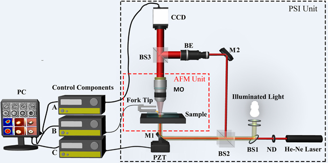
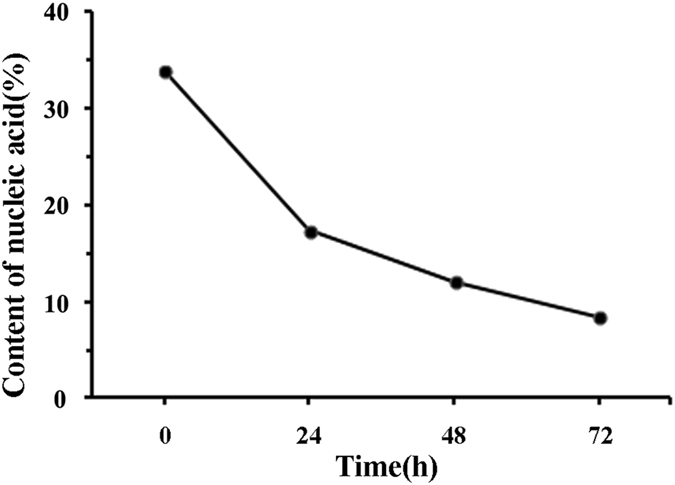
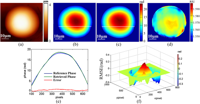
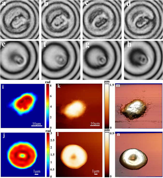
Cell refractive index, an intrinsic optical parameter, is closely correlated with the intracellular mass and concentration. By combining optical phase-shifting interferometry (PSI) and atomic force microscope (AFM) imaging, we constructed a label free, non-invasive and quantitative refractive index of single cell measurement system, in which the accurate phase map of single cell was retrieved with PSI technique and the cell morphology with nanoscale resolution was achieved with AFM imaging. Based on the proposed AFM/PSI system, we achieved quantitative refractive index distributions of single red blood cell and Jurkat cell, respectively. Further, the quantitative change of refractive index distribution during Daunorubicin (DNR)-induced Jurkat cell apoptosis was presented, and then the content changes of intracellular biochemical components were achieved. Importantly, these results were consistent with Raman spectral analysis, indicating that the proposed PSI/AFM based refractive index system is likely to become a useful tool for intracellular biochemical components analysis measurement, and this will facilitate its application for revealing cell structure and pathological state from a new perspective.
细胞折射率是一个固有光学参数,与细胞内质量和浓度密切相关。通过结合光学相移干涉测量(PSI)和原子力显微镜(AFM)成像,我们构建了一种无标记、非侵入性和定量的单细胞折射率测量系统,该系统利用 PSI 技术获取单细胞的精确相位图,并利用 AFM 成像获得具有纳米级分辨率的细胞形态。基于所提出的 AFM/PSI 系统,我们分别实现了单个红细胞和 Jurkat 细胞的定量折射率分布。进一步,展示了柔红霉素(DNR)诱导 Jurkat 细胞凋亡过程中折射率分布的定量变化,进而实现了细胞内生化成分含量的变化。重要的是,这些结果与拉曼光谱分析一致,表明所提出的基于 PSI/AFM 的折射率系统很可能成为细胞内生化成分分析测量的有用工具,这将有助于从新的角度揭示细胞结构和病理状态。