• 文献检索
  • 文档翻译
  • 深度研究
  • 学术资讯
  • Suppr Zotero 插件Zotero 插件
  • 邀请有礼
  • 套餐&价格
  • 历史记录
应用&插件
Suppr Zotero 插件Zotero 插件浏览器插件Mac 客户端Windows 客户端微信小程序
定价
高级版会员购买积分包购买API积分包
服务
文献检索文档翻译深度研究API 文档MCP 服务
关于我们
关于 Suppr公司介绍联系我们用户协议隐私条款
关注我们

Suppr 超能文献

核心技术专利:CN118964589B侵权必究
粤ICP备2023148730 号-1Suppr @ 2026

文献检索

告别复杂PubMed语法,用中文像聊天一样搜索,搜遍4000万医学文献。AI智能推荐,让科研检索更轻松。

立即免费搜索

文件翻译

保留排版,准确专业,支持PDF/Word/PPT等文件格式,支持 12+语言互译。

免费翻译文档

深度研究

AI帮你快速写综述,25分钟生成高质量综述,智能提取关键信息,辅助科研写作。

立即免费体验

除基底细胞癌外的各种皮肤病的皮肤镜下树枝状血管

Arborizing Vessels on Dermoscopy in Various Skin Diseases Other Than Basal Cell Carcinoma.

作者信息

Jin Hyunju, Yang Min-Young, Kim Jeong-Min, Kim Gun-Wook, Kim Hoon-Soo, Ko Hyun-Chang, Kim Byung-Soo, Kim Moon-Bum

机构信息

Department of Dermatology, Pusan National University School of Medicine, Yangsan, Korea.

Department of Dermatology, Pusan National University Yangsan Hospital, Yangsan, Korea.

出版信息

Ann Dermatol. 2017 Jun;29(3):288-294. doi: 10.5021/ad.2017.29.3.288. Epub 2017 May 11.

DOI:10.5021/ad.2017.29.3.288
PMID:28566904
原文链接:https://pmc.ncbi.nlm.nih.gov/articles/PMC5438934/
Abstract

BACKGROUND

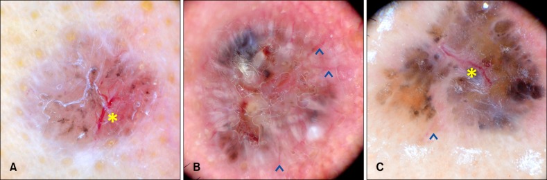
Arborizing vessels (AVs) are dermoscopically defined as telangiectasias with distinct treelike branching, and are a characteristic feature of basal cell carcinoma (BCC). However, AVs are observed in various conditions other than BCC.

OBJECTIVE

The aim of this study was to investigate skin diseases showing AV and investigates dermoscopic differences between BCC and non-BCC.

METHODS

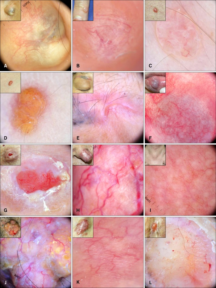
Dermoscopic images showing AV were prospectively collected and classified into BCC/non-BCC. Non-BCC was further classified into tumors (benign cystic, benign non-cystic, premalignant, and malignant) and non-tumors. We compared AV focusing, widest diameter of stem vessels, widest diameter ratio of stem vessel to first branch, and number of ramifications between groups.

RESULTS

Among 124 images, 54.0% were BCC and 46.0% were non-BCC. Non-BCC included epidermal cysts, hypertrophic scars/keloids, intradermal nevi, actinic keratoses, etc. The proportion of focused AV in BCC was significantly higher and the proportion of unfocused AV in BCC was lower than that of premalignant and malignant non-BCC. The widest diameter ratio of stem vessel to first branch was higher in non-BCC. Number of ramifications was significantly less in benign cystic non-BCC than BCC.

CONCLUSION

Various skin diseases showed AV, so that diagnoses other than BCC should be considered. The findings in this study could help discriminate BCC from non-BCC.

摘要

背景

树枝状血管(AVs)在皮肤镜下被定义为具有明显树状分支的毛细血管扩张,是基底细胞癌(BCC)的特征性表现。然而,除BCC外,在多种情况下也可观察到AVs。

目的

本研究旨在调查显示AVs的皮肤疾病,并研究BCC与非BCC之间的皮肤镜差异。

方法

前瞻性收集显示AVs的皮肤镜图像,并分为BCC/非BCC。非BCC进一步分为肿瘤(良性囊性、良性非囊性、癌前病变和恶性)和非肿瘤。我们比较了各组之间AVs的聚焦情况、主干血管的最宽直径、主干血管与第一分支的最宽直径比以及分支数量。

结果

在124张图像中,54.0%为BCC,46.0%为非BCC。非BCC包括表皮囊肿、肥厚性瘢痕/瘢痕疙瘩、皮内痣、光化性角化病等。BCC中聚焦AVs的比例显著高于癌前病变和恶性非BCC,而BCC中未聚焦AVs的比例低于癌前病变和恶性非BCC。非BCC中主干血管与第一分支的最宽直径比更高。良性囊性非BCC的分支数量显著少于BCC。

结论

多种皮肤疾病可显示AVs,因此应考虑除BCC以外的诊断。本研究结果有助于鉴别BCC与非BCC。

https://cdn.ncbi.nlm.nih.gov/pmc/blobs/6095/5438934/91d28421435d/ad-29-288-g002.jpg
https://cdn.ncbi.nlm.nih.gov/pmc/blobs/6095/5438934/a0a1fb345e5d/ad-29-288-g001.jpg
https://cdn.ncbi.nlm.nih.gov/pmc/blobs/6095/5438934/91d28421435d/ad-29-288-g002.jpg
https://cdn.ncbi.nlm.nih.gov/pmc/blobs/6095/5438934/a0a1fb345e5d/ad-29-288-g001.jpg
https://cdn.ncbi.nlm.nih.gov/pmc/blobs/6095/5438934/91d28421435d/ad-29-288-g002.jpg

相似文献

1
Arborizing Vessels on Dermoscopy in Various Skin Diseases Other Than Basal Cell Carcinoma.除基底细胞癌外的各种皮肤病的皮肤镜下树枝状血管
Ann Dermatol. 2017 Jun;29(3):288-294. doi: 10.5021/ad.2017.29.3.288. Epub 2017 May 11.
2
Dermoscopic features of basal cell carcinoma and its subtypes: A systematic review.基底细胞癌及其亚型的皮肤镜特征:系统综述。
J Am Acad Dermatol. 2021 Sep;85(3):653-664. doi: 10.1016/j.jaad.2019.11.008. Epub 2019 Nov 7.
3
Dermoscopic Clues of Histopathologically Aggressive Basal Cell Carcinoma Subtypes.组织病理侵袭性基底细胞癌亚型的皮肤镜线索。
Medicina (Kaunas). 2023 Feb 13;59(2):349. doi: 10.3390/medicina59020349.
4
A multicentric study on dermoscopic patterns and clinical-dermoscopic-histological correlates of basal cell carcinoma in Indian skin.一项关于印度皮肤基底细胞癌的皮肤镜模式和临床-皮肤镜-组织学相关性的多中心研究。
Clin Exp Dermatol. 2022 Nov;47(11):1982-1990. doi: 10.1111/ced.15337. Epub 2022 Oct 9.
5
Clinical, Dermoscopic and Histopathological Evaluation of Basal Cell Carcinoma.基底细胞癌的临床、皮肤镜及组织病理学评估
Dermatol Pract Concept. 2023 Jan 1;13(1):e2023004. doi: 10.5826/dpc.1301a4.
6
Basal Cell Carcinoma Mimicking Desmoplastic Trichoepithelioma: A Case with Correlation of Dermoscopy and Histology.酷似促结缔组织增生性毛发上皮瘤的基底细胞癌:1例皮肤镜与组织学对照病例
Case Rep Dermatol. 2018 May 18;10(2):133-137. doi: 10.1159/000489164. eCollection 2018 May-Aug.
7
Statistical evaluation of dermoscopic features in basal cell carcinomas.基底细胞癌皮肤镜特征的统计学评估
Dermatol Surg. 2014 Jul;40(7):718-24. doi: 10.1111/dsu.0000000000000031.
8
Accuracy of dermoscopic criteria for discriminating superficial from other subtypes of basal cell carcinoma.基于皮肤镜特征鉴别基底细胞癌表浅型与其他亚型的准确性。
J Am Acad Dermatol. 2014 Feb;70(2):303-11. doi: 10.1016/j.jaad.2013.10.003. Epub 2013 Nov 20.
9
Dermoscopic Features of Basal Cell Carcinoma on the Lower Limbs: A Chameleon!下肢基底细胞癌的皮肤镜特征:变色龙!
Dermatology. 2017;233(6):482-488. doi: 10.1159/000487300. Epub 2018 Mar 22.
10
Dermoscopy of skin lesions in two patients with xeroderma pigmentosum.两名着色性干皮病患者皮肤病变的皮肤镜检查
Br J Dermatol. 2005 Feb;152(2):271-8. doi: 10.1111/j.1365-2133.2004.06332.x.

引用本文的文献

1
A Rare Case of Male Breast Cancer Presenting as a Superficial Pigmented Nipple Tumor: The Importance of a Deep Excisional Biopsy and Immunohistochemical Analysis in Diagnosis.一例表现为浅表色素沉着乳头肿瘤的男性乳腺癌罕见病例:深部切除活检及免疫组化分析在诊断中的重要性
Cureus. 2025 May 2;17(5):e83353. doi: 10.7759/cureus.83353. eCollection 2025 May.
2
Unsealed Rhenium Resin Brachytherapy in Non-Surgical Candidates With Refractory Basal Cell Carcinoma: Clinical Outcomes.未密封铼树脂近距离放射治疗非手术候选的难治性基底细胞癌:临床结果
Dermatol Pract Concept. 2025 Apr 1;15(2):4993. doi: 10.5826/dpc.1502a4933.
3
Dermoscopy of Basal Cell Carcinoma Part 3: Differential Diagnosis, Treatment Monitoring and Novel Technologies.

本文引用的文献

1
A pearly papule with arborizing vessels.一个伴有树枝状血管的珍珠样丘疹。
J Am Acad Dermatol. 2015 Jan;72(1 Suppl):S25-6. doi: 10.1016/j.jaad.2014.04.058.
2
Dermatoscopic pattern of digital mucous cyst: report of three cases.数字黏液囊肿的皮肤镜模式:三例报告。
Dermatol Pract Concept. 2014 Oct 31;4(4):65-7. doi: 10.5826/dpc.0404a12. eCollection 2014 Oct.
3
Keloids and hypertrophic scars: characteristic vascular structures visualized by using dermoscopy.瘢痕疙瘩和增生性瘢痕:通过皮肤镜观察到的特征性血管结构
基底细胞癌的皮肤镜检查 第3部分:鉴别诊断、治疗监测及新技术
Cancers (Basel). 2025 Mar 19;17(6):1025. doi: 10.3390/cancers17061025.
4
Giant Morpheaform Basal Cell Carcinoma Mimicking Scarring Alopecia: Exception Prone to Neglect.酷似瘢痕性脱发的巨大苔藓样基底细胞癌:易被忽视的罕见病例
Dermatopathology (Basel). 2024 Jun 5;11(2):154-160. doi: 10.3390/dermatopathology11020016.
Ann Dermatol. 2014 Oct;26(5):603-9. doi: 10.5021/ad.2014.26.5.603. Epub 2014 Sep 26.
4
Dermoscopy of basosquamous carcinoma.基底鳞状细胞癌的皮肤镜检查。
Br J Dermatol. 2013 Aug;169(2):358-64. doi: 10.1111/bjd.12394.
5
Arborizing vessels under dermoscopy: a case of cellular neurothekeoma instead of basal cell carcinoma.皮肤镜下呈树枝状血管:一例细胞性神经鞘瘤而非基底细胞癌
Dermatol Online J. 2013 Mar 15;19(3):5.
6
Dermoscopy of dermatofibrosarcoma protuberans: a study of 15 cases.隆突性皮肤纤维肉瘤的皮肤镜检查:15 例研究。
Br J Dermatol. 2013 Jul;169(1):85-90. doi: 10.1111/bjd.12318.
7
Dermoscopy of desmoplastic trichoepithelioma reveals other criteria to distinguish it from basal cell carcinoma.皮肤镜检查有助于区分隆凸性毛囊角化病和基底细胞癌。
Dermatology. 2013;226(2):101-4. doi: 10.1159/000346246. Epub 2013 Jan 30.
8
Dermoscopy of apocrine hidrocystomas: a morphological study.顶泌汗腺汗管瘤的皮肤镜检查:形态学研究。
J Eur Acad Dermatol Venereol. 2014 Mar;28(3):378-81. doi: 10.1111/jdv.12044. Epub 2012 Dec 1.
9
Dermoscopy findings of pseudolymphomatous folliculitis.假性淋巴瘤性毛囊炎的皮肤镜表现
Case Rep Dermatol. 2012 May;4(2):154-7. doi: 10.1159/000341194. Epub 2012 Jul 12.
10
Dermoscopy of Merkel cell carcinoma.默克尔细胞癌的皮肤镜检查。
Dermatology. 2012;224(2):140-4. doi: 10.1159/000337411. Epub 2012 Apr 6.