Dermatology Unit, Department of Clinical Medicine and Surgery, University of Naples Federico II, 80131 Naples, Italy.
Department of Dermatology, "Iuliu Hațieganu" University of Medicine and Pharmacy, 400012 Cluj-Napoca, Romania.
Medicina (Kaunas). 2023 Feb 13;59(2):349. doi: 10.3390/medicina59020349.
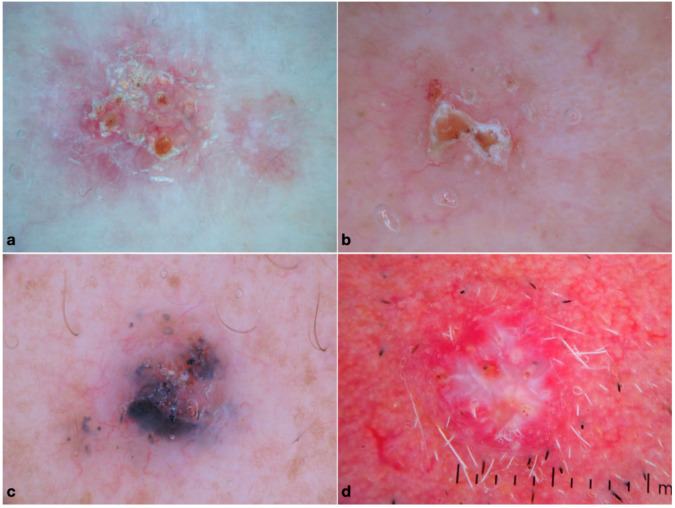
The group of histopathologically aggressive BCC subtypes includes morpheaform, micronodular, infiltrative and metatypical BCC. Since these tumors are at increased risk of recurring, micrographically controlled surgery is considered the best therapeutic option. Although dermoscopy significantly improves the clinical recognition of BCC, scarce evidence exists on their dermoscopic criteria. To investigate the dermoscopic characteristics of histopathologically aggressive BCC subtypes. Dermoscopic images of morpheaform, micronodular, infiltrative and metatypical BCC were analyzed for the presence of predefined variables. Descriptive and analytical statistics were performed. Most histopathologically aggressive BCCs were located on the head and neck. Infiltrative was the most common subtype. All subtypes, except micronodular BCC, rarely displayed dermoscopic pigmentation. The most frequent dermoscopic features of infiltrative BCC were arborizing vessels (67.1%), shiny white structures (48.6%) and ulceration (52.9%). The features prevailing in morpheaform BCC were arborizing vessels (68.4%), ulceration ( = 12, 63.2%) and white porcelain areas (47.4%). Micronodular BCC was typified by milky red structureless areas (53.8%), arborizing vessels (53.8%), short fine telangiectasias (50%), ulceration (46.2%) and blue structures (57.7%). The most common findings in metatypical BCC were arborizing vessels (77.8%), shiny white structures (66.7%), ulceration (62.9%) and keratin mass (29.6%). Study population of only white skin and relatively small sample size in some groups. Our study provided data on the clinical, dermoscopic and epidemiological characteristics of histopathologically aggressive BCCs.
组织学上侵袭性基底细胞癌亚型包括硬斑病样型、微结节型、浸润型和 型。由于这些肿瘤复发风险增加,显微镜下控制的手术被认为是最佳治疗选择。虽然皮肤镜显著提高了基底细胞癌的临床识别率,但关于其皮肤镜标准的证据很少。 探讨组织学侵袭性基底细胞癌亚型的皮肤镜特征。 对硬斑病样型、微结节型、浸润型和 型基底细胞癌的皮肤镜图像进行了分析,以确定是否存在预定的变量。进行了描述性和分析性统计。 大多数组织学侵袭性基底细胞癌位于头颈部。浸润型是最常见的亚型。除微结节型基底细胞癌外,所有亚型很少显示皮肤镜色素沉着。浸润性基底细胞癌最常见的皮肤镜特征是树枝状血管(67.1%)、闪亮的白色结构(48.6%)和溃疡(52.9%)。硬斑病样型基底细胞癌的特征是树枝状血管(68.4%)、溃疡(12 例,63.2%)和白色瓷状区域(47.4%)。微结节型基底细胞癌的特征是乳白色无结构区域(53.8%)、树枝状血管(53.8%)、短而细的毛细血管扩张(50%)、溃疡(46.2%)和蓝色结构(57.7%)。 型基底细胞癌最常见的发现是树枝状血管(77.8%)、闪亮的白色结构(66.7%)、溃疡(62.9%)和角蛋白团块(29.6%)。 仅研究了白种人群,且某些组的样本量较小。 我们的研究提供了组织学侵袭性基底细胞癌的临床、皮肤镜和流行病学特征数据。