Pino Jessica M V, da Luz Marcio H M, Antunes Hanna K M, Giampá Sara Q de Campos, Martins Vilma R, Lee Kil S
Departamento de Bioquímica, Universidade Federal de São PauloSão Paulo, Brazil.
Departamento de Psicobiologia, Universidade Federal de São PauloSão Paulo, Brazil.
Front Mol Neurosci. 2017 May 17;10:145. doi: 10.3389/fnmol.2017.00145. eCollection 2017.
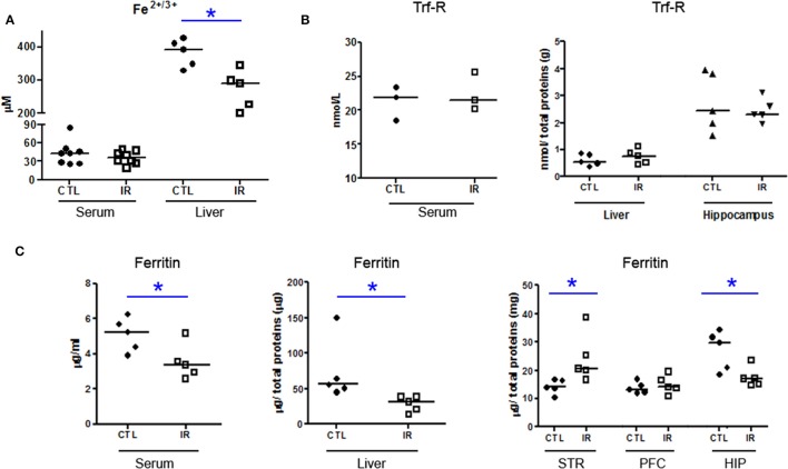
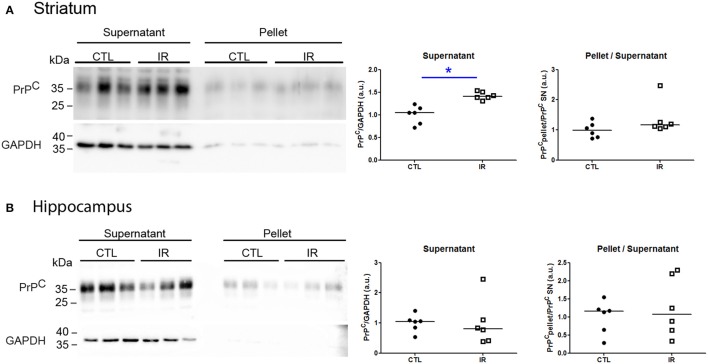
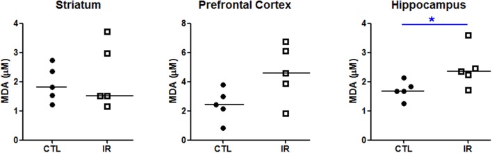
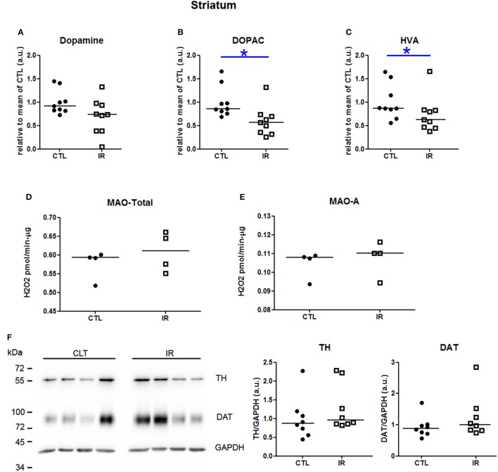
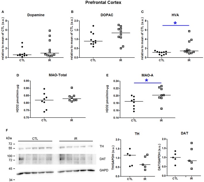
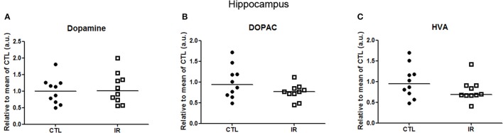
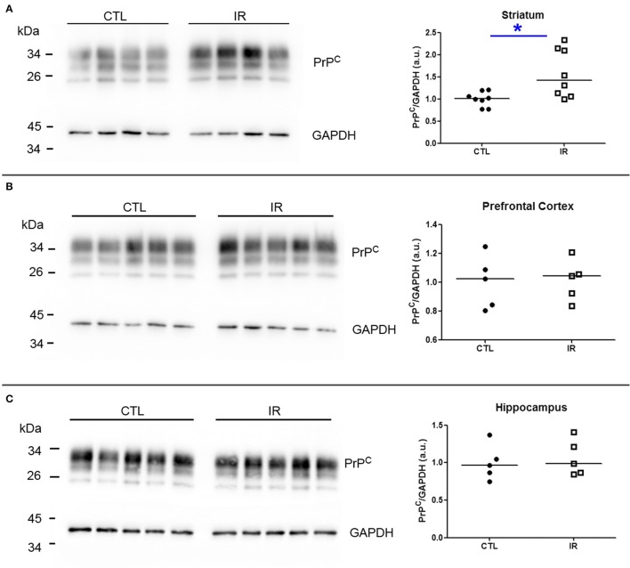
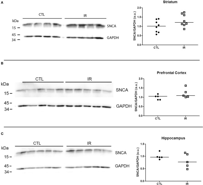
Iron is an essential micronutrient for several physiological functions, including the regulation of dopaminergic neurotransmission. On the other hand, both iron, and dopamine can affect the folding and aggregation of proteins related with neurodegenerative diseases, such as cellular prion protein (PrP) and α-synuclein, suggesting that deregulation of iron homeostasis and the consequential disturbance of dopamine metabolism can be a risk factor for conformational diseases. These proteins, in turn, are known to participate in the regulation of iron and dopamine metabolism. In this study, we evaluated the effects of dietary iron restriction on brain ferritin levels, dopamine metabolism, and the expression levels of PrP and α-synuclein. To achieve this goal, C57BL/6 mice were fed with iron restricted diet (IR) or with normal diet (CTL) for 1 month. IR reduced iron and ferritin levels in liver. Ferritin reduction was also observed in the hippocampus. However, in the striatum of IR group, ferritin level was increased, suggesting that under iron-deficient condition, each brain area might acquire distinct capacity to store iron. Increased lipid peroxidation was observed only in hippocampus of IR group, where ferritin level was reduced. IR also generated discrete results regarding dopamine metabolism of distinct brain regions: in striatum, the level of dopamine metabolites (DOPAC and HVA) was reduced; in prefrontal cortex, only HVA was increased along with the enhanced MAO-A activity; in hippocampus, no alterations were observed. PrP levels were increased only in the striatum of IR group, where ferritin level was also increased. PrP is known to play roles in iron uptake. Thus, the increase of PrP in striatum of IR group might be related to the increased ferritin level. α-synuclein was not altered in any regions. Abnormal accumulation of ferritin, increased MAO-A activity or lipid peroxidation are molecular features observed in several neurological disorders. Our findings show that nutritional iron deficiency produces these molecular alterations in a region-specific manner and provide new insight into the variety of molecular pathways that can lead to distinct neurological symptoms upon iron deficiency. Thus, adequate iron supplementation is essential for brain health and prevention of neurological diseases.
铁是多种生理功能所必需的微量营养素,包括对多巴胺能神经传递的调节。另一方面,铁和多巴胺都会影响与神经退行性疾病相关的蛋白质的折叠和聚集,如细胞朊蛋白(PrP)和α-突触核蛋白,这表明铁稳态失调以及随之而来的多巴胺代谢紊乱可能是构象性疾病的一个风险因素。反过来,这些蛋白质已知参与铁和多巴胺代谢的调节。在本研究中,我们评估了饮食中铁限制对脑铁蛋白水平、多巴胺代谢以及PrP和α-突触核蛋白表达水平的影响。为实现这一目标,将C57BL/6小鼠喂食铁限制饮食(IR)或正常饮食(CTL)1个月。IR降低了肝脏中的铁和铁蛋白水平。在海马体中也观察到铁蛋白减少。然而,在IR组的纹状体中,铁蛋白水平升高,这表明在缺铁条件下,每个脑区可能具有不同的铁储存能力。仅在IR组铁蛋白水平降低的海马体中观察到脂质过氧化增加。IR对不同脑区的多巴胺代谢也产生了不同的结果:在纹状体中,多巴胺代谢产物(DOPAC和HVA)水平降低;在额叶前皮质中,只有HVA随着MAO-A活性增强而增加;在海马体中,未观察到变化。PrP水平仅在IR组铁蛋白水平也升高的纹状体中增加。已知PrP在铁摄取中起作用。因此,IR组纹状体中PrP的增加可能与铁蛋白水平升高有关。α-突触核蛋白在任何区域均未改变。铁蛋白异常积累、MAO-A活性增加或脂质过氧化是在几种神经系统疾病中观察到的分子特征。我们的研究结果表明,营养性缺铁以区域特异性方式产生这些分子改变,并为缺铁时可导致不同神经症状的多种分子途径提供了新的见解。因此,充足的铁补充对于脑健康和预防神经系统疾病至关重要。