Oh Joo Han, Kim Woo, Cayetano Angel A
Department of Orthopedic Surgery, Seoul National University College of Medicine, Seoul National University Bundang Hospital, Seongnam, Korea.
Department of Orthopaedic Surgery, Nalgae Hospital, Seoul, Korea.
Clin Orthop Surg. 2017 Jun;9(2):223-231. doi: 10.4055/cios.2017.9.2.223. Epub 2017 May 8.
Humeral retroversion is variable among individuals, and there are several measurement methods. This study was conducted to compare the concordance and reliability between the standard method and 5 other measurement methods on two-dimensional (2D) computed tomography (CT) scans.
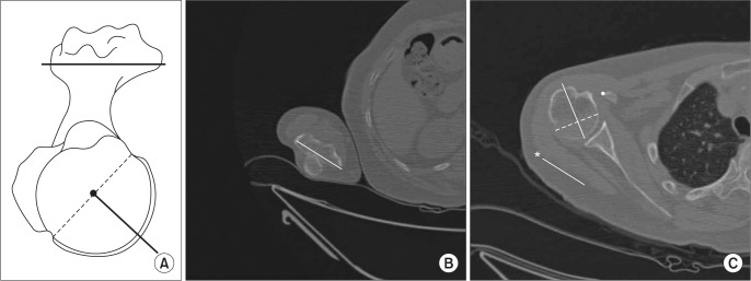
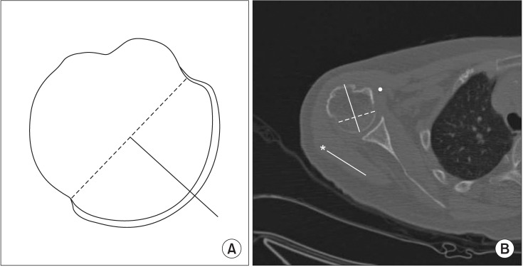
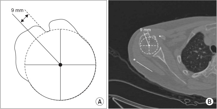
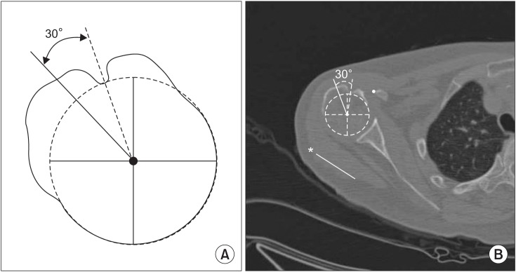
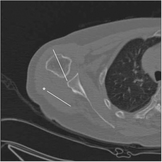
CT scans from 21 patients who underwent shoulder arthroplasty (19 women and 2 men; mean age, 70.1 years [range, 42 to 81 years]) were analyzed. The elbow transepicondylar axis was used as a distal reference. Proximal reference points included the central humeral head axis (standard method), the axis of the humeral center to 9 mm posterior to the posterior margin of the bicipital groove (method 1), the central axis of the bicipital groove -30° (method 2), the base axis of the triangular shaped metaphysis +2.5° (method 3), the distal humeral head central axis +2.4° (method 4), and contralateral humeral head retroversion (method 5). Measurements were conducted independently by two orthopedic surgeons.
The mean humeral retroversion was 31.42° ± 12.10° using the standard method, and 29.70° ± 11.66° (method 1), 30.64° ± 11.24° (method 2), 30.41° ± 11.17° (method 3), 32.14° ± 11.70° (method 4), and 34.15° ± 11.47° (method 5) for the other methods. Interobserver reliability and intraobserver reliability exceeded 0.75 for all methods. On the test to evaluate the equality of the standard method to the other methods, the intraclass correlation coefficients (ICCs) of method 2 and method 4 were different from the ICC of the standard method in surgeon A ( < 0.05), and the ICCs of method 2 and method 3 were different form the ICC of the standard method in surgeon B ( < 0.05).
Humeral version measurement using the posterior margin of the bicipital groove (method 1) would be most concordant with the standard method even though all 5 methods showed excellent agreements.
肱骨扭转角度在个体间存在差异,且有多种测量方法。本研究旨在比较二维(2D)计算机断层扫描(CT)上标准方法与其他5种测量方法之间的一致性和可靠性。
分析了21例行肩关节置换术患者的CT扫描数据(19名女性和2名男性;平均年龄70.1岁[范围42至81岁])。以肘关节髁上轴作为远端参考。近端参考点包括肱骨头中心轴(标准方法)、肱骨头中心至肱二头肌沟后缘后方9 mm处的轴线(方法1)、肱二头肌沟中心轴-30°(方法2)、三角形干骺端基部轴+2.5°(方法3)、肱骨头远端中心轴+2.4°(方法4)以及对侧肱骨头扭转角度(方法5)。由两名骨科医生独立进行测量。
使用标准方法测得的平均肱骨扭转角度为31.42°±12.10°,其他方法分别为:方法1为29.70°±11.66°,方法2为30.64°±11.24°,方法3为30.41°±11.17°,方法4为32.14°±11.70°,方法5为34.15°±11.47°。所有方法的观察者间可靠性和观察者内可靠性均超过0.75。在评估标准方法与其他方法一致性的测试中,方法2和方法4的组内相关系数(ICC)在外科医生A中与标准方法的ICC不同(<0.05),方法2和方法3的ICC在外科医生B中与标准方法的ICC不同(<0.05)。
尽管所有5种方法都显示出极好的一致性,但使用肱二头肌沟后缘进行肱骨扭转角度测量(方法1)与标准方法最为一致。