Wang Mengyan, Tang Feng, Pan Xiaobo, Yao Longfang, Wang Xinyi, Jing Yueyue, Ma Jiong, Wang Guifang, Mi Lan
Department of Optical Science and Engineering, Shanghai Engineering Research Center of Ultra-Precision Optical Manufacturing, Key Laboratory of Micro and Nano Photonic Structures (Ministry of Education), Green Photoelectron Platform, Fudan University, 220 Handan Road, Shanghai 200433, China.
Department of Pathology, Huashan Hospital, Fudan University, 12 Wulumuqi Middle Road, Shanghai 200040, China.
BBA Clin. 2017 Apr 27;8:7-13. doi: 10.1016/j.bbacli.2017.04.002. eCollection 2017 Dec.
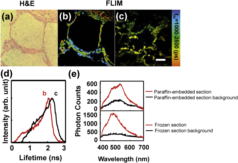
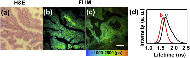
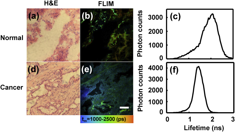
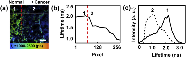
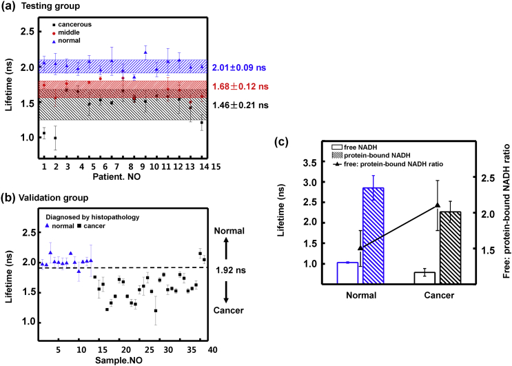
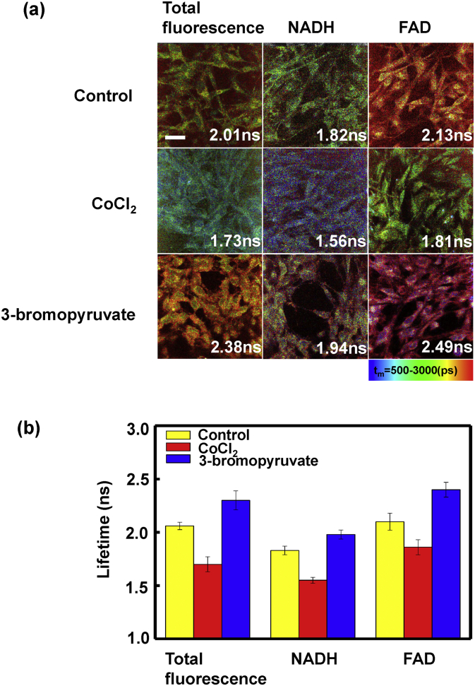
A method of rapidly differentiating lung tumor from healthy tissue is extraordinarily needed for both the diagnosis and the intraoperative margin assessment. We assessed the ability of fluorescence lifetime imaging microscopy (FLIM) for differentiating human lung cancer and normal tissues with the autofluorescence, and also elucidated the mechanism in tissue studies and cell studies. A 15-patient testing group was used to compare FLIM results with traditional histopathology diagnosis. Based on the endogenous fluorescence lifetimes of the testing group, a criterion line was proposed to distinguish normal and cancerous tissues. Then by blinded examined 41 sections from the validation group of other 16 patients, the sensitivity and specificity of FLIM were determined. The cellular metabolism was studied with specific perturbations of oxidative phosphorylation and glycolysis in cell studies. The fluorescence lifetime of cancerous lung tissues is consistently lower than normal tissues, and this is due to the both decrease of reduced nicotinamide adenine dinucleotide (NADH) and flavin adenine dinucleotide (FAD) lifetimes. A criterion line of lifetime at 1920 ps can be given for differentiating human lung cancer and normal tissues.The sensitivity and specificity of FLIM for lung cancer diagnosis were determined as 92.9% and 92.3%. These findings suggest that NADH and FAD can be used to rapidly diagnose lung cancer. FLIM is a rapid, accurate and highly sensitive technique in the judgment during lung cancer surgery and it can be potential in earlier cancer detection.
无论是对于肺癌的诊断还是术中切缘评估,都极其需要一种能够快速区分肺肿瘤与健康组织的方法。我们评估了荧光寿命成像显微镜(FLIM)利用自发荧光区分人类肺癌组织和正常组织的能力,并在组织研究和细胞研究中阐明了其机制。使用一个由15名患者组成的测试组,将FLIM结果与传统组织病理学诊断结果进行比较。基于测试组的内源性荧光寿命,提出了一条区分正常组织和癌组织的标准线。然后,通过对另外16名患者的验证组的41个切片进行盲法检查,确定了FLIM的敏感性和特异性。在细胞研究中,通过对氧化磷酸化和糖酵解进行特定扰动来研究细胞代谢。癌性肺组织的荧光寿命始终低于正常组织,这是由于还原型烟酰胺腺嘌呤二核苷酸(NADH)和黄素腺嘌呤二核苷酸(FAD)的寿命均降低所致。可以给出一条1920 ps的寿命标准线来区分人类肺癌组织和正常组织。FLIM对肺癌诊断的敏感性和特异性分别确定为92.9%和92.3%。这些发现表明,NADH和FAD可用于快速诊断肺癌。FLIM在肺癌手术判断中是一种快速、准确且高度灵敏的技术,在早期癌症检测中具有潜力。