Suda Kenichi, Rozeboom Leslie, Rivard Christopher J, Yu Hui, Ellison Kim, Melnick Mary Ann C, Hinz Trista K, Chan Daniel, Heasley Lynn E, Politi Katerina, Mitsudomi Tetsuya, Hirsch Fred R
Division of Medical Oncology, University of Colorado Anschutz Medical Campus, 12801 E. 17th Ave, RC-1 South, Aurora, CO 80045, USA; Division of Thoracic Surgery, Department of Surgery, Kindai University Faculty of Medicine, 377-2 Ohno-higashi, Osaka-Sayama 589-0014, Japan.
Division of Medical Oncology, University of Colorado Anschutz Medical Campus, 12801 E. 17th Ave, RC-1 South, Aurora, CO 80045, USA.
Lung Cancer. 2017 Jul;109:1-8. doi: 10.1016/j.lungcan.2017.04.010. Epub 2017 Apr 19.
Immunotherapy that targets the programmed death-1/programmed death-ligand 1 (PD-L1) axis has been approved for treatment of non-small cell lung cancer (NSCLC) patients in many countries. However, our current understanding of the role of immunotherapies on NSCLC patients with epidermal growth factor receptor (EGFR) mutation, following acquisition of resistance to EGFR tyrosine kinase inhibitors (TKIs), is so far unclear. Especially, there is little data on if each acquired resistance mechanism to EGFR-TKIs alters PD-L1 expression status which is employed as an important predictive biomarker for PD-1/PD-L1 targeting agents.
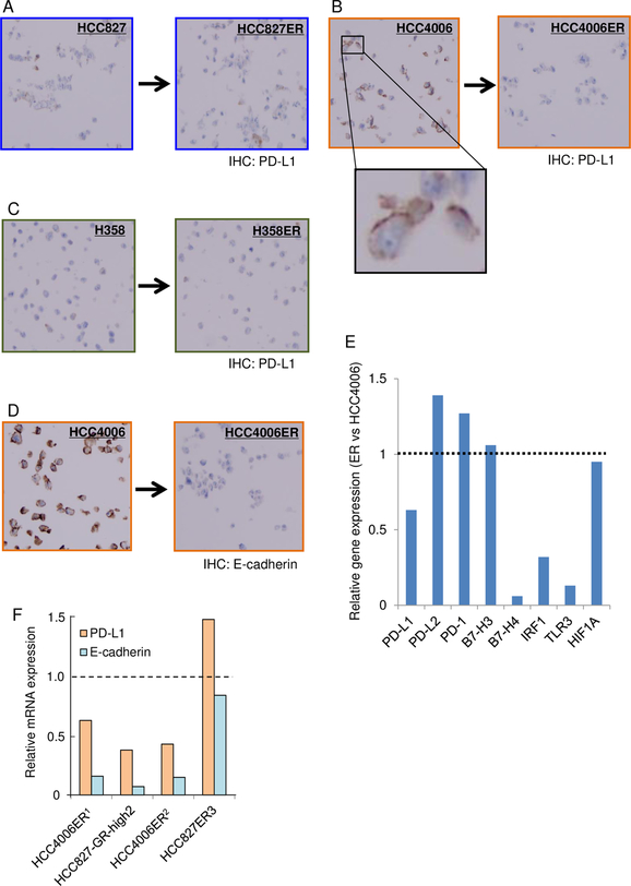
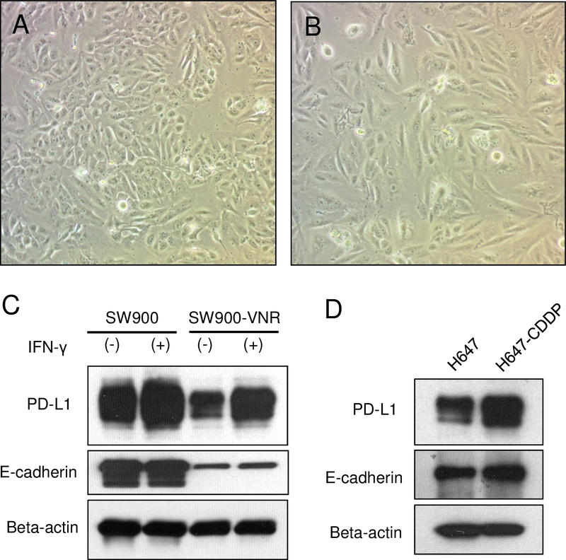
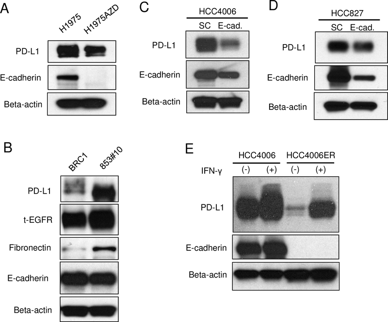
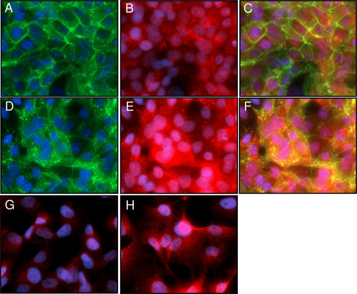
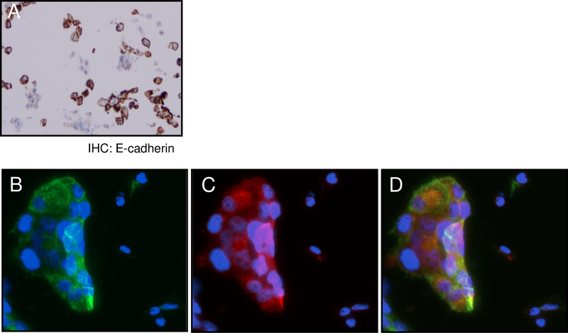
Lung cancer cell lines (HCC827, HCC4006, PC9, H1975, H358, SW900, and H647) and their daughter cells that acquired resistance to EGFR-TKIs or cytotoxic drugs (cisplatin or vinorelbine) were examined. PD-L1 expression was analyzed by immunohistochemistry, immunoblotting, and/or fluorescent imaging. Published microarray data were also employed to evaluate our findings.
We found correlations between therapy-induced E-cadherin downregulation and decreased PD-L1 expression using our cell lines and published microarray data. ShRNA mediated E-cadherin knockdown decreased PD-L1 expression in parental cells, and dual immunofluorescent staining of E-cadherin and PD-L1 suggests co-localization of both molecules. We also observed marked downregulation of PD-L1 in cells with E-cadherin downregulation after chronic treatment with vinorelbine. These results indicate a correlation between therapy-induced E-cadherin downregulation and decreased PD-L1 expression, highlighting the importance of re-biopsy after acquisition of resistance to EGFR-TKIs, not only for the evaluation of resistance mechanisms but also for the determination of PD-L1 expression status.
靶向程序性死亡蛋白1/程序性死亡配体1(PD-L1)轴的免疫疗法已在许多国家被批准用于治疗非小细胞肺癌(NSCLC)患者。然而,目前我们对免疫疗法在表皮生长因子受体(EGFR)突变的NSCLC患者中,继对EGFR酪氨酸激酶抑制剂(TKIs)产生耐药性之后所起作用的了解尚不清楚。特别是,关于EGFR-TKIs的每种获得性耐药机制是否会改变PD-L1表达状态的数据很少,而PD-L1表达状态是PD-1/PD-L1靶向药物的重要预测生物标志物。
检测肺癌细胞系(HCC827、HCC4006、PC9、H1975、H358、SW900和H647)及其对EGFR-TKIs或细胞毒性药物(顺铂或长春瑞滨)产生耐药性的子代细胞。通过免疫组织化学、免疫印迹和/或荧光成像分析PD-L1表达。还利用已发表的微阵列数据来评估我们的研究结果。
利用我们的细胞系和已发表的微阵列数据,我们发现治疗诱导的E-钙黏蛋白下调与PD-L1表达降低之间存在相关性。短发夹RNA(shRNA)介导的E-钙黏蛋白敲低降低了亲本细胞中的PD-L1表达,并且E-钙黏蛋白和PD-L1的双重免疫荧光染色表明这两种分子共定位。在用长春瑞滨长期处理后,我们还观察到E-钙黏蛋白下调的细胞中PD-L1明显下调。这些结果表明治疗诱导的E-钙黏蛋白下调与PD-L1表达降低之间存在相关性,突出了在对EGFR-TKIs产生耐药性后重新活检的重要性,这不仅用于评估耐药机制,还用于确定PD-L1表达状态。