Malejko Kathrin, Abler Birgit, Plener Paul L, Straub Joana
Department of Psychiatry and Psychotherapy III, University Hospital Ulm, Ulm, Germany.
Department of Child and Adolescent Psychiatry and Psychotherapy, University Hospital Ulm, Ulm, Germany.
Front Psychiatry. 2017 May 19;8:85. doi: 10.3389/fpsyt.2017.00085. eCollection 2017.
Post-traumatic stress disorder (PTSD) is a common psychiatric disease with changes in neural circuitries. Neurobiological models conceptualize the symptoms of PTSD as correlates of a dysfunctional stress reaction to traumatic events. Functional imaging studies showed an increased amygdala and a decreased prefrontal cortex response in PTSD patients. As psychotherapeutic approaches represent the gold standard for PTSD treatment, it is important to examine its underlying neurobiological correlates.
Studies published until August 2016 were selected through systematic literature research in the databases PubMed, PsychInfo, and Cochrane Library's Central Register of Controlled Trials or were identified manually by searching reference lists of selected articles. Search terms were "neural correlates" OR "fMRI" OR "SPECT," AND "therapy" AND "PTSD." A total of 19 articles were included in the present review whereof 15 studies compared pre-to-post-therapy signal changes, six studies related pre-treatment activity to pre-to-post-symptom improvement, and four studies compared neural correlates of responders versus non-responders. The disposed therapy forms were cognitive behavioral therapy (CBT), eye movement desensitization and reprocessing, cognitive therapy, exposure therapy, mindfulness-based intervention, brief eclectic psychotherapy, and unspecified therapy.
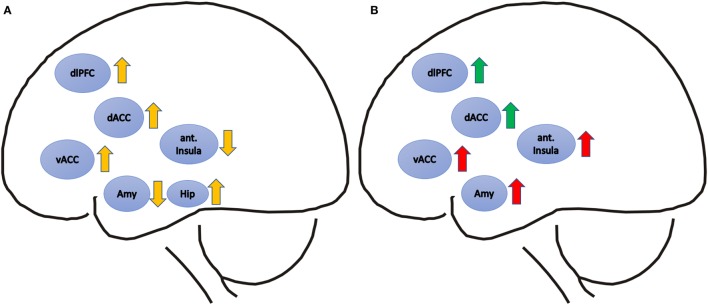
Successful psychotherapy of PTSD was repeatedly shown to be accompanied by decreased activity in the amygdala and the insula as well as increased activity in the dorsal anterior cingulate cortex (dACC) and hippocampus. Elevated dACC activity prior to treatment was related to subsequent treatment success and a positive predictor for treatment response. Elevated amygdala and insula pre-treatment activities were related to treatment failure.
Decreased activity in limbic brain regions and increased activity in frontal brain areas in PTSD patients after successful psychotherapeutic treatment might reflect regained top-down control over previously impaired bottom-up processes.
创伤后应激障碍(PTSD)是一种常见的精神疾病,伴有神经回路的改变。神经生物学模型将PTSD的症状概念化为对创伤事件功能失调的应激反应的相关因素。功能成像研究显示,PTSD患者杏仁核活性增加,前额叶皮质反应降低。由于心理治疗方法是PTSD治疗的金标准,因此研究其潜在的神经生物学相关性很重要。
通过在PubMed、PsychInfo和Cochrane图书馆对照试验中央注册库中进行系统文献检索,选择截至2016年8月发表的研究,或通过搜索所选文章的参考文献列表手动识别。搜索词为“神经相关性”或“功能磁共振成像”或“单光子发射计算机断层扫描”,以及“治疗”和“创伤后应激障碍”。本综述共纳入19篇文章,其中15项研究比较了治疗前后的信号变化,6项研究将治疗前的活动与症状改善前后的情况相关联,4项研究比较了反应者与无反应者的神经相关性。所采用的治疗形式包括认知行为疗法(CBT)、眼动脱敏再处理疗法、认知疗法、暴露疗法、基于正念的干预、简短折衷心理治疗和未明确说明的疗法。
PTSD的成功心理治疗反复显示伴随着杏仁核和脑岛活动的减少以及背侧前扣带回皮质(dACC)和海马体活动的增加。治疗前dACC活性升高与随后的治疗成功相关,是治疗反应的积极预测指标。治疗前杏仁核和脑岛活性升高与治疗失败相关。
PTSD患者在成功的心理治疗后,边缘脑区活动减少,额叶脑区活动增加,这可能反映了对先前受损的自下而上过程重新获得了自上而下的控制。