• 文献检索
  • 文档翻译
  • 深度研究
  • 学术资讯
  • Suppr Zotero 插件Zotero 插件
  • 邀请有礼
  • 套餐&价格
  • 历史记录
应用&插件
Suppr Zotero 插件Zotero 插件浏览器插件Mac 客户端Windows 客户端微信小程序
定价
高级版会员购买积分包购买API积分包
服务
文献检索文档翻译深度研究API 文档MCP 服务
关于我们
关于 Suppr公司介绍联系我们用户协议隐私条款
关注我们

Suppr 超能文献

核心技术专利:CN118964589B侵权必究
粤ICP备2023148730 号-1Suppr @ 2026

文献检索

告别复杂PubMed语法,用中文像聊天一样搜索,搜遍4000万医学文献。AI智能推荐,让科研检索更轻松。

立即免费搜索

文件翻译

保留排版,准确专业,支持PDF/Word/PPT等文件格式,支持 12+语言互译。

免费翻译文档

深度研究

AI帮你快速写综述,25分钟生成高质量综述,智能提取关键信息,辅助科研写作。

立即免费体验

心脏磁共振容积-时间曲线评估2型糖尿病合并冠状动脉疾病患者的左心室功能障碍

Volume-time curve of cardiac magnetic resonance assessed left ventricular dysfunction in coronary artery disease patients with type 2 diabetes mellitus.

作者信息

Xu Hua-Yan, Yang Zhi-Gang, Guo Ying-Kun, Shi Ke, Liu Xi, Zhang Qin, Jiang Li, Xie Lin-Jun

机构信息

Department of Radiology, National Key Laboratory of Biotherapy, West China Hospital, Sichuan University, # 37Guo Xue Xiang, Chengdu, Sichuan, 610041, China.

Department of Radiology, West China Second University Hospital, Sichuan University, #20, Section 3, Renmin South Road, Chengdu, Sichuan, 610041, China.

出版信息

BMC Cardiovasc Disord. 2017 Jun 5;17(1):145. doi: 10.1186/s12872-017-0583-5.

DOI:10.1186/s12872-017-0583-5
PMID:28583071
原文链接:https://pmc.ncbi.nlm.nih.gov/articles/PMC5460487/
Abstract

BACKGROUND

Type 2 diabetes mellitus (DM2) may induce epicardial coronary artery diseases and left ventricular myocardial damaging as well. Left ventricular dysfunction was found in DM2. In this research, we compared the left ventricular dysfunction of coronary artery disease (CAD) patients with and without type 2 diabetes mellitus as well as normal controls using the volume-time curve of cardiac magnetic resonance (CMR).

METHODS

Sixty-one CAD patients (28 with DM2 and 33 without DM2) and 18 normal individuals were enrolled in this study. Left ventricular function parameters, including the end-diastolic and end-systolic volumes (EDV, ESV), stroke volume (SV) and ejection fraction (EF), and morphologic dimension parameters (end diastolic and systolic diameter (EDD and ESD), were measured and compared. Volume-time curve parameters, including the peak ejection rate (PER), peak ejection time (PET), peak filling rate (PFR), peak filling time from ES (PFT), peak ejection rate normalized to EDV (PER/EDV), and peak filling rate normalized to EDV (PFR/EDV), were derived automatically and compared.

RESULTS

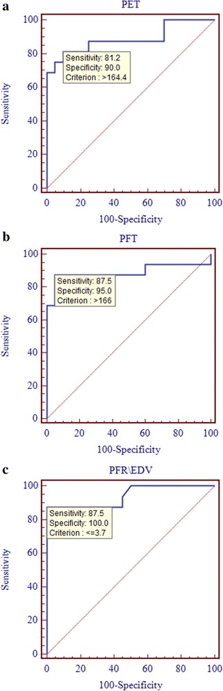
LVEF in the diabetic CAD group was markedly reduced when compared to the normal and CAD without DM2 groups (all p < 0.05). LVEDD of the diabetic CAD group was significantly enlarged compared to the normal and non-diabetic CAD groups (all p < 0.05). More importantly, the lowest parameters of the left ventricle volume time curve (i.e., PER, PFR, PER/EDV and PFR/EDV) were obtained in diabetic CAD patients (all p < 0.05). In diabetic CAD patients, logistic regression analysis indicated that PET, PFT and PFR/EDV were independent predictors of left ventricular dysfunction (odds ratio [OR]: 1.1208, 1.0161, and 0.0139, respectively). The sensitivity and specificity of PET were 81.2 and 90%, respectively, when the threshold value was greater than 164.4 msec; for PFT, the sensitivity and specificity were 87.5 and 95.0%, respectively (criterion >166.0 msec). Higher sensitivity (87.5%) and specificity (100.0%) were obtained for PFR/EDV (criterion ≤3.7EDV/s).

CONCLUSIONS

Parameters that are derived from the volume-time curve on CMR, including PET, PFT and PFR/EDV, allow clinicians to predict left ventricular dysfunction in diabetic CAD subjects with a high degree of sensitivity and specificity.

摘要

背景

2型糖尿病(DM2)可能诱发心外膜冠状动脉疾病以及左心室心肌损伤。在DM2患者中发现了左心室功能障碍。在本研究中,我们使用心脏磁共振成像(CMR)的容积-时间曲线,比较了合并和不合并2型糖尿病的冠心病(CAD)患者以及正常对照者的左心室功能障碍情况。

方法

本研究纳入了61例CAD患者(28例合并DM2,33例不合并DM2)和18名正常个体。测量并比较左心室功能参数,包括舒张末期和收缩末期容积(EDV、ESV)、每搏输出量(SV)和射血分数(EF),以及形态学尺寸参数(舒张末期和收缩末期直径(EDD和ESD))。自动得出并比较容积-时间曲线参数,包括峰值射血率(PER)、峰值射血时间(PET)、峰值充盈率(PFR)、从收缩末期开始的峰值充盈时间(PFT)、以EDV标准化的峰值射血率(PER/EDV)以及以EDV标准化的峰值充盈率(PFR/EDV)。

结果

与正常组和不合并DM2的CAD组相比,糖尿病合并CAD组的左心室射血分数(LVEF)显著降低(所有p<0.05)。与正常组和非糖尿病CAD组相比,糖尿病合并CAD组的左心室舒张末期内径(LVEDD)显著增大(所有p<0.05)。更重要的是,糖尿病合并CAD患者的左心室容积-时间曲线参数最低(即PER、PFR、PER/EDV和PFR/EDV)(所有p<0.05)。在糖尿病合并CAD患者中,逻辑回归分析表明PET、PFT和PFR/EDV是左心室功能障碍的独立预测因素(优势比[OR]分别为1.1208、1.0161和0.0139)。当阈值大于164.4毫秒时,PET的敏感性和特异性分别为81.2%和90%;对于PFT,敏感性和特异性分别为87.5%和95.0%(标准>166.0毫秒)。PFR/EDV的敏感性(87.5%)和特异性(100.0%)更高(标准≤3.7EDV/s)。

结论

CMR容积-时间曲线上得出的参数,包括PET、PFT和PFR/EDV,使临床医生能够以高度的敏感性和特异性预测糖尿病合并CAD患者的左心室功能障碍。

https://cdn.ncbi.nlm.nih.gov/pmc/blobs/3a4c/5460487/07df24ff6b35/12872_2017_583_Fig4_HTML.jpg
https://cdn.ncbi.nlm.nih.gov/pmc/blobs/3a4c/5460487/0ebeace936cc/12872_2017_583_Fig1_HTML.jpg
https://cdn.ncbi.nlm.nih.gov/pmc/blobs/3a4c/5460487/75dd1c6f1220/12872_2017_583_Fig2_HTML.jpg
https://cdn.ncbi.nlm.nih.gov/pmc/blobs/3a4c/5460487/3ec8218e6b14/12872_2017_583_Fig3_HTML.jpg
https://cdn.ncbi.nlm.nih.gov/pmc/blobs/3a4c/5460487/07df24ff6b35/12872_2017_583_Fig4_HTML.jpg

相似文献

1
Volume-time curve of cardiac magnetic resonance assessed left ventricular dysfunction in coronary artery disease patients with type 2 diabetes mellitus.心脏磁共振容积-时间曲线评估2型糖尿病合并冠状动脉疾病患者的左心室功能障碍
BMC Cardiovasc Disord. 2017 Jun 5;17(1):145. doi: 10.1186/s12872-017-0583-5.
2
Impact of type 2 diabetes mellitus on left ventricular diastolic function in patients with essential hypertension: evaluation by volume-time curve of cardiac magnetic resonance.2 型糖尿病对原发性高血压患者左心室舒张功能的影响:心脏磁共振容积-时间曲线的评估。
Cardiovasc Diabetol. 2021 Mar 25;20(1):73. doi: 10.1186/s12933-021-01262-1.
3
Left ventricular diastolic function in type 2 diabetes mellitus and the association with coronary artery calcium score: a cardiac MRI study.2 型糖尿病患者的左心室舒张功能与冠状动脉钙评分的关系:一项心脏 MRI 研究。
AJR Am J Roentgenol. 2014 Jun;202(6):1207-14. doi: 10.2214/AJR.13.11325.
4
Diastolic dysfunction assessed by ultra-fast cadmium-zinc-telluride cardiac imaging: impact on the evaluation of ischaemia.超快速碲化镉锌心脏成像评估的舒张功能障碍:对缺血评估的影响。
Eur Heart J Cardiovasc Imaging. 2015 Jan;16(1):68-73. doi: 10.1093/ehjci/jeu166. Epub 2014 Sep 3.
5
[Comparison of left and right ventricular ejection and filling parameters of the heart using cine-MRI with breath holding technique. Clinical study of 42 patients with cardiomyopathy and coronary heart disease].[采用屏气技术的电影磁共振成像比较心脏左右心室射血及充盈参数。42例心肌病和冠心病患者的临床研究]
Rofo. 1999 Jun;170(6):534-41. doi: 10.1055/s-2007-1011088.
6
Magnetic resonance assessment of left ventricular diastolic dysfunction for detecting cardiac allograft vasculopathy in recipients of heart transplants.心脏移植受者左心室舒张功能障碍的磁共振评估检测心脏移植后冠状动脉病。
Int J Cardiovasc Imaging. 2012 Mar;28(3):555-62. doi: 10.1007/s10554-011-9853-y. Epub 2011 Mar 26.
7
Evaluation of elevated left ventricular end diastolic pressure in patients with preserved ejection fraction using cardiac magnetic resonance.应用心脏磁共振评估射血分数保留的心力衰竭患者的左心室舒张末期压力升高。
Eur Radiol. 2019 May;29(5):2360-2368. doi: 10.1007/s00330-018-5955-4. Epub 2019 Jan 10.
8
Adenosine stress CMR T1-mapping detects early microvascular dysfunction in patients with type 2 diabetes mellitus without obstructive coronary artery disease.腺苷负荷心脏磁共振 T1 mapping 检测 2 型糖尿病无阻塞性冠状动脉疾病患者的早期微血管功能障碍。
J Cardiovasc Magn Reson. 2017 Oct 25;19(1):81. doi: 10.1186/s12968-017-0397-8.
9
Reproducibility of peak filling and peak emptying rate determined by cardiovascular magnetic resonance imaging for assessment of biventricular systolic and diastolic dysfunction in patients with pulmonary arterial hypertension.通过心血管磁共振成像测定的峰值充盈率和峰值排空率在评估肺动脉高压患者双心室收缩和舒张功能障碍中的可重复性。
Int J Cardiovasc Imaging. 2018 May;34(5):777-786. doi: 10.1007/s10554-017-1281-1. Epub 2017 Nov 22.
10
Wall stress determines systolic and diastolic function--Characteristics of heart failure.壁应力决定收缩和舒张功能——心力衰竭的特征。
Int J Cardiol. 2016 Jan 1;202:685-93. doi: 10.1016/j.ijcard.2015.09.032. Epub 2015 Sep 25.

引用本文的文献

1
Association of the triglyceride‒glucose index with subclinical right ventricular systolic dysfunction in type 2 diabetes mellitus patients.2型糖尿病患者甘油三酯-葡萄糖指数与亚临床右心室收缩功能障碍的关联
Sci Rep. 2025 Jun 6;15(1):19915. doi: 10.1038/s41598-025-05356-8.
2
Diastolic dysfunction in adults with uncomplicated obesity evaluated with left atrial and left ventricular tissue tracking and ventricular volume-time curve: a prospective cardiac magnetic resonance study.采用左心房和左心室组织追踪及心室容积-时间曲线评估单纯性肥胖成人的舒张功能障碍:一项前瞻性心脏磁共振研究
Quant Imaging Med Surg. 2024 Jul 1;14(7):5040-5056. doi: 10.21037/qims-23-1785. Epub 2024 Jun 13.
3

本文引用的文献

1
Coronary artery disease in type 2 diabetes mellitus: Recent treatment strategies and future perspectives.2型糖尿病中的冠状动脉疾病:近期治疗策略与未来展望
World J Cardiol. 2015 Mar 26;7(3):119-24. doi: 10.4330/wjc.v7.i3.119.
2
Relation of late gadolinium enhancement in cardiac magnetic resonance on the diastolic volume recovery of left ventricle with hypertrophic cardiomyopathy.心脏磁共振钆延迟增强与肥厚型心肌病左心室舒张末期容积恢复的关系。
J Thorac Dis. 2014 Jul;6(7):988-94. doi: 10.3978/j.issn.2072-1439.2014.06.37.
3
Left ventricular diastolic function in type 2 diabetes mellitus and the association with coronary artery calcium score: a cardiac MRI study.
Integration of longitudinal and circumferential strain predicts volumetric change across the cardiac cycle and differentiates patients along the heart failure continuum.
纵向应变和周向应变的整合可预测整个心动周期的容积变化,并沿心力衰竭连续统区分患者。
J Cardiovasc Magn Reson. 2023 Oct 2;25(1):55. doi: 10.1186/s12968-023-00969-2.
4
Reference data for left ventricular filling and atrial function in children using cardiovascular magnetic resonance.应用心血管磁共振成像评估儿童左心室充盈和心房功能的参考数据
J Cardiovasc Magn Reson. 2023 Jun 12;25(1):30. doi: 10.1186/s12968-023-00936-x.
5
Early longitudinal changes in left ventricular function and morphology in diabetic pigs: evaluation by 3.0T magnetic resonance imaging.3.0T 磁共振成像评价糖尿病猪左心室功能和形态的早期纵向变化。
Cardiovasc Diabetol. 2023 Jan 10;22(1):6. doi: 10.1186/s12933-022-01734-y.
6
Cardiac magnetic resonance diastolic indices correlate with ventricular filling pressures in pediatric heart transplant recipients.心脏磁共振舒张指数与儿科心脏移植受者的心室充盈压相关。
Pediatr Transplant. 2022 Sep;26(6):e14332. doi: 10.1111/petr.14332. Epub 2022 Jun 10.
7
Effects of selective heart rate reduction with ivabradine on LV function and central hemodynamics in patients with chronic coronary syndrome.伊伐布雷定选择性降低心率对慢性冠状动脉综合征患者左心室功能和中心血流动力学的影响。
Int J Cardiol Heart Vasc. 2021 Mar 23;34:100757. doi: 10.1016/j.ijcha.2021.100757. eCollection 2021 Jun.
8
Impact of type 2 diabetes mellitus on left ventricular diastolic function in patients with essential hypertension: evaluation by volume-time curve of cardiac magnetic resonance.2 型糖尿病对原发性高血压患者左心室舒张功能的影响:心脏磁共振容积-时间曲线的评估。
Cardiovasc Diabetol. 2021 Mar 25;20(1):73. doi: 10.1186/s12933-021-01262-1.
9
Cardiac magnetic resonance feature tracking for quantifying right ventricular deformation in type 2 diabetes mellitus patients.心脏磁共振特征追踪技术定量评估 2 型糖尿病患者右心室变形。
Sci Rep. 2019 Jul 31;9(1):11148. doi: 10.1038/s41598-019-46755-y.
2 型糖尿病患者的左心室舒张功能与冠状动脉钙评分的关系:一项心脏 MRI 研究。
AJR Am J Roentgenol. 2014 Jun;202(6):1207-14. doi: 10.2214/AJR.13.11325.
4
2013 ESC guidelines on the management of stable coronary artery disease: the Task Force on the management of stable coronary artery disease of the European Society of Cardiology.2013年欧洲心脏病学会稳定型冠状动脉疾病管理指南:欧洲心脏病学会稳定型冠状动脉疾病管理特别工作组
Eur Heart J. 2013 Oct;34(38):2949-3003. doi: 10.1093/eurheartj/eht296. Epub 2013 Aug 30.
5
Advances in parametric mapping with CMR imaging.CMR 成像的参数图研究进展。
JACC Cardiovasc Imaging. 2013 Jul;6(7):806-22. doi: 10.1016/j.jcmg.2013.05.005.
6
Diabetes and coronary heart disease: a risk factor for the global epidemic.糖尿病与冠心病:全球流行病的一个风险因素。
Int J Hypertens. 2012;2012:697240. doi: 10.1155/2012/697240. Epub 2012 Oct 18.
7
Screening for coronary artery disease in patients with type 2 diabetes mellitus: An evidence-based review.2型糖尿病患者冠状动脉疾病的筛查:一项基于证据的综述。
Indian J Endocrinol Metab. 2012 Jan;16(1):94-101. doi: 10.4103/2230-8210.91202.
8
Left ventricular diastolic function in type 2 diabetes mellitus is associated with myocardial triglyceride content but not with impaired myocardial perfusion reserve.2 型糖尿病患者的左心室舒张功能与心肌甘油三酯含量有关,但与心肌灌注储备受损无关。
J Magn Reson Imaging. 2012 Apr;35(4):804-11. doi: 10.1002/jmri.22879. Epub 2011 Nov 8.
9
Left ventricular filling patterns in patients with previous myocardial infarction measured by conventional cine cardiac magnetic resonance.应用传统电影心脏磁共振测量陈旧性心肌梗死患者的左心室充盈模式。
Int J Cardiovasc Imaging. 2012 Apr;28(4):795-801. doi: 10.1007/s10554-011-9883-5. Epub 2011 May 7.
10
Assessment of left ventricular function: visual or quantitative?左心室功能评估:视觉评估还是定量评估?
Int J Cardiovasc Imaging. 2011 Apr;27(4):573-7. doi: 10.1007/s10554-010-9735-8. Epub 2010 Oct 28.