Department of Pharmacology and Clinical Neuroscience, Umeå University, Umeå, Sweden.
BMC Pharmacol Toxicol. 2017 Jun 5;18(1):42. doi: 10.1186/s40360-017-0151-8.
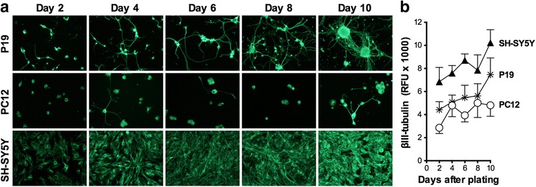
Exposure to chemicals might be toxic to the developing brain. There is a need for simple and robust in vitro cellular models for evaluation of chemical-induced neurotoxicity as a complement to traditional studies on animals. In this study, neuronally differentiated mouse embryonal carcinoma P19 cells (P19 neurons) were compared with human neuroblastoma SH-SY5Y cells and rat adrenal pheochromocytoma PC12 cells for their ability to detect toxicity of methylmercury (MeHg), okadaic acid and acrylamide.
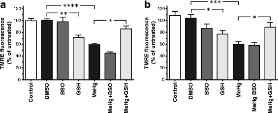
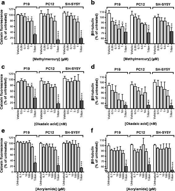
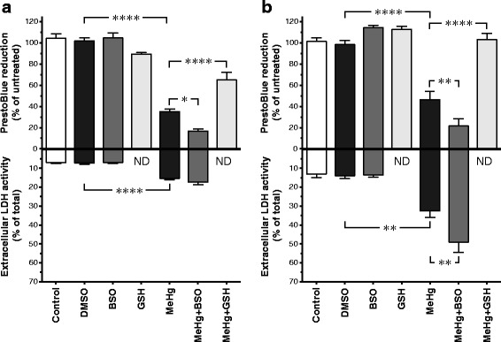
Retinoic acid-treated P19 and SH-SY5Y cells and nerve growth factor-stimulated PC12 cells, allowed to differentiate for 6 days, were exposed to MeHg, okadaic acid and acrylamide for 48 h. Cell survival and neurite outgrowth were assessed with the calcein-AM assay and fluorescence detection of antibodies against the cytoskeletal neuron-specific protein βIII-tubulin, respectively. The effects of glutathione (GSH) and the potent inhibitor of GSH synthesis buthionine sulfoximine (BSO) on the MeHg induced-toxicity were assessed using the PrestoBlue™ cell viability assay and the TMRE mitochondrial membrane potential assay.
Differentiated P19 cells developed the most extensive neuronal network among the three cell models and were the most sensitive neuronal model to detect neurotoxic effects of the test compounds. MeHg produced a concentration-dependent toxicity in differentiated P19 cells and SH-SY5Y cells, with statistically significant effects at concentrations from 0.1 μM in the P19 neurons and 1 μM in the SH-SY5Y cells. MeHg induced a decrease in the cellular metabolic activity and mitochondrial membrane potential (ΔΨm) in the differentiated P19 cells and SH-SY5Y cells, that were attenuated by GSH. Okadaic acid and acrylamide also showed statistically significant toxicity in the P19 neurons, but not in the SH-SY5Y cells or the P12 cells.
P19 neurons are more sensitive to detect cytotoxicity of MeHg, okadaic acid and acrylamide than retinoic acid-differentiated SH-SY5Y cells and nerve growth factor-treated PC12 cells. P19 neurons are at least as sensitive as differentiated SH-SY5Y cells to detect the loss of mitochondrial membrane potential produced by MeHg and the protective effects of extracellular GSH on MeHg toxicity. P19 neurons may be a useful model to study neurotoxic effects of chemicals.
接触化学物质可能对发育中的大脑有毒。因此,需要建立简单而强大的体外细胞模型来评估化学诱导的神经毒性,以作为传统动物研究的补充。在这项研究中,我们比较了神经分化的小鼠胚胎癌细胞 P19 细胞(P19 神经元)与人类神经母细胞瘤 SH-SY5Y 细胞和大鼠肾上腺嗜铬细胞瘤 PC12 细胞,以评估其对甲基汞(MeHg)、岗田酸和丙烯酰胺毒性的检测能力。
用视黄酸处理 P19 和 SH-SY5Y 细胞,并给予神经生长因子刺激 PC12 细胞分化 6 天,然后用 MeHg、岗田酸和丙烯酰胺处理 48 小时。通过 calcein-AM 测定法评估细胞存活率,通过荧光检测抗细胞骨架神经元特异性蛋白 βIII-微管蛋白的抗体评估神经突生长。使用 PrestoBlueTM 细胞活力测定法和 TMRE 线粒体膜电位测定法评估谷胱甘肽 (GSH) 和强效 GSH 合成抑制剂丁硫氨酸亚砜胺 (BSO) 对 MeHg 诱导毒性的影响。
分化的 P19 细胞在三种细胞模型中形成了最广泛的神经元网络,并且是检测测试化合物神经毒性作用的最敏感的神经元模型。MeHg 在分化的 P19 细胞和 SH-SY5Y 细胞中产生浓度依赖性毒性,在 P19 神经元中浓度为 0.1μM,在 SH-SY5Y 细胞中浓度为 1μM 时具有统计学意义。MeHg 诱导分化的 P19 细胞和 SH-SY5Y 细胞的细胞代谢活性和线粒体膜电位(ΔΨm)下降,而 GSH 可减弱这种下降。岗田酸和丙烯酰胺在 P19 神经元中也表现出统计学显著的毒性,但在 SH-SY5Y 细胞或 PC12 细胞中则没有。
与视黄酸分化的 SH-SY5Y 细胞和神经生长因子处理的 PC12 细胞相比,P19 神经元对 MeHg、岗田酸和丙烯酰胺的细胞毒性检测更敏感。P19 神经元对 MeHg 引起的线粒体膜电位丧失和细胞外 GSH 对 MeHg 毒性的保护作用的检测敏感性至少与分化的 SH-SY5Y 细胞相同。P19 神经元可能是研究化学物质神经毒性作用的有用模型。