Wu Yaoqun, Zhang Pei, Yang Hongyun, Ge Yong, Xin Yong
Department of Dermatology, Xiangyang Hospital, Hubei University of Medicine, Xiangyang, Hubei 441000, P.R. China.
Department of Radiotherapy, Affiliated Hospital of Xuzhou Medical University, Xuzhou, Jiangsu 221002, P.R. China.
Mol Med Rep. 2017 Jul;16(1):539-546. doi: 10.3892/mmr.2017.6666. Epub 2017 May 31.
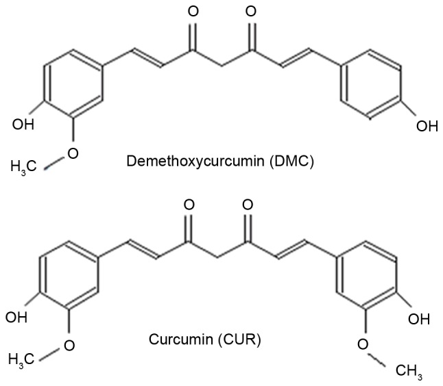
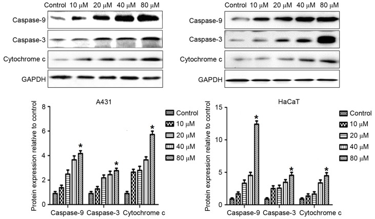
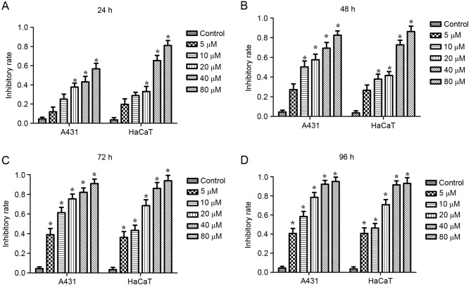
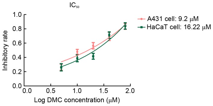
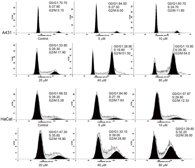
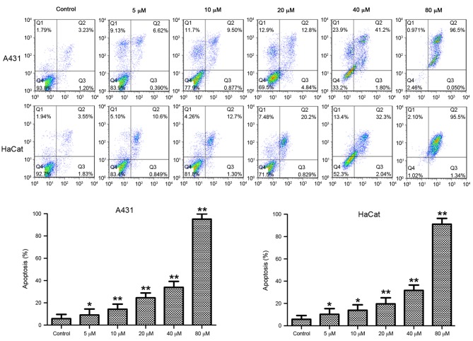
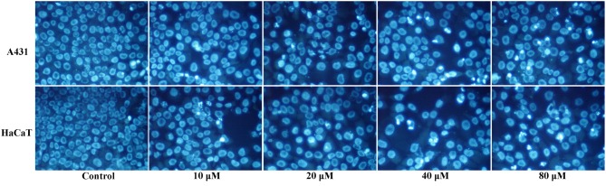
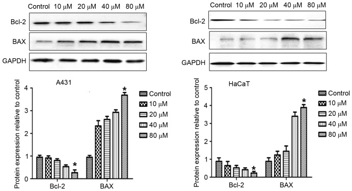
The present study investigated the effects and mechanisms of demethoxycurcumin (DMC) on a human skin squamous cell carcinoma cell line, A431, and a human keratinocyte cell line, HaCaT. A431 and HaCaT cells were cultured in vitro. The effects of DMC treatment on cell viability were analyzed using the Cell Counting kit‑8 (CCK‑8) assay; cell cycle distribution was analyzed by flow cytometry; apoptosis was assessed by flow cytometry and Hoechst 33258 staining; and the protein expression levels of cytochrome c, B‑cell lymphoma 2 (Bcl‑2), Bcl‑2‑associated X protein (BAX), caspase‑9 and caspase‑3 were evaluated by western blotting. CCK‑8 assay results demonstrated that DMC treatment significantly inhibited viability of A431 and HaCaT cells in a dose‑dependent manner. Flow cytometric analysis confirmed that DMC treatment induced apoptosis in a dose‑dependent manner, and significantly increased the proportion of cells in G2/M phase. Western blot analysis indicated that the protein expression levels of Bcl‑2 were decreased, whereas the expression levels of BAX, caspase‑9, caspase‑3 and cytochrome c were increased following DMC treatment compared with in untreated cells. In conclusion, DMC treatment significantly inhibited viability of A431 and HaCaT cells, and induced cell cycle arrest in G2/M phase. The present study indicated that DMC may induce apoptosis of skin cancer cells through a caspase‑dependent pathway.
本研究调查了去甲氧基姜黄素(DMC)对人皮肤鳞状细胞癌细胞系A431和人角质形成细胞系HaCaT的作用及其机制。A431和HaCaT细胞在体外培养。使用细胞计数试剂盒-8(CCK-8)分析法分析DMC处理对细胞活力的影响;通过流式细胞术分析细胞周期分布;通过流式细胞术和Hoechst 33258染色评估细胞凋亡;并通过蛋白质印迹法评估细胞色素c、B细胞淋巴瘤2(Bcl-2)、Bcl-2相关X蛋白(BAX)、半胱天冬酶-9和半胱天冬酶-3的蛋白质表达水平。CCK-8分析结果表明,DMC处理以剂量依赖性方式显著抑制A431和HaCaT细胞的活力。流式细胞术分析证实,DMC处理以剂量依赖性方式诱导细胞凋亡,并显著增加G2/M期细胞的比例。蛋白质印迹分析表明,与未处理的细胞相比,DMC处理后Bcl-2的蛋白质表达水平降低,而BAX、半胱天冬酶-9、半胱天冬酶-3和细胞色素c的表达水平升高。总之,DMC处理显著抑制A431和HaCaT细胞的活力,并诱导细胞周期停滞在G2/M期。本研究表明,DMC可能通过半胱天冬酶依赖性途径诱导皮肤癌细胞凋亡。