Tien Thomas, Zhang Joyce, Muto Tetsuya, Kim Dongjoon, Sarthy Vijay P, Roy Sayon
Departments of Medicine and Ophthalmology, Boston University School of Medicine, Boston, Massachusetts, United States.
Department of Ophthalmology, Northwestern University Feinberg School of Medicine, Chicago, Illinois, United States.
Invest Ophthalmol Vis Sci. 2017 Jun 1;58(7):2915-2921. doi: 10.1167/iovs.16-21355.
To investigate whether high glucose (HG) induces mitochondrial dysfunction and promotes apoptosis in retinal Müller cells.
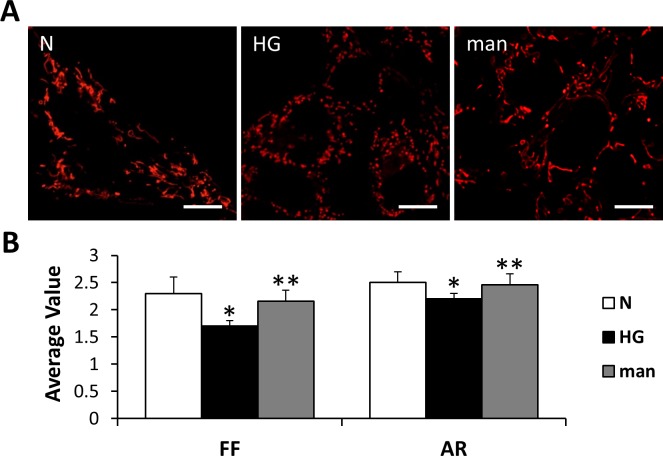
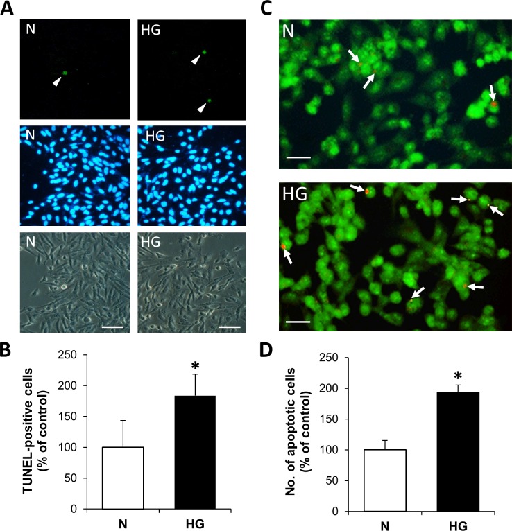
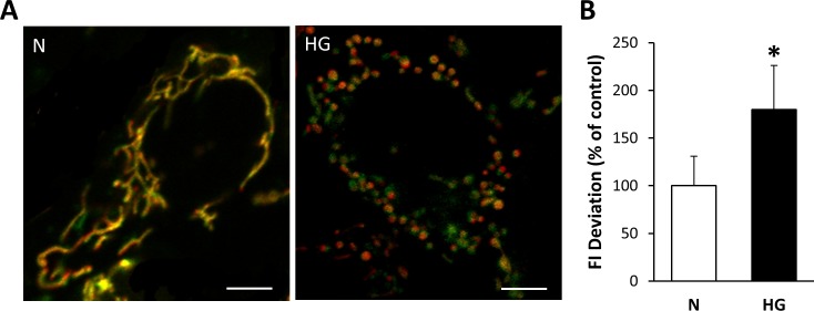
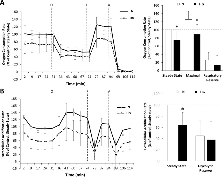
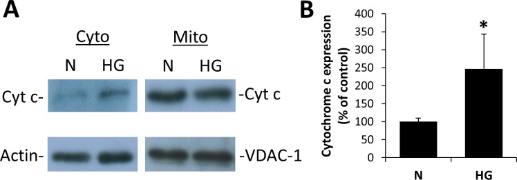
Rat retinal Müller cells (rMC-1) grown in normal (N) or HG (30 mM glucose) medium for 7 days were subjected to MitoTracker Red staining to identify the mitochondrial network. Digital images of mitochondria were captured in live cells under confocal microscopy and analyzed for mitochondrial morphology changes based on form factor (FF) and aspect ratio (AR) values. Mitochondrial metabolic function was assessed by measuring oxygen consumption rate (OCR) and extracellular acidification rate (ECAR) using a bioenergetic analyzer. Cells undergoing apoptosis were identified by differential dye staining and TUNEL assay, and cytochrome c levels were assessed by Western blot analysis.
Cells grown in HG exhibited significantly increased mitochondrial fragmentation compared to those grown in N medium (FF = 1.7 ± 0.1 vs. 2.3 ± 0.1; AR = 2.1 ± 0.1 vs. 2.5 ± 0.2; P < 0.01). OCR and ECAR were significantly reduced in cells grown in HG medium compared to those grown in N medium (steady state: 75% ± 20% of control, P < 0.02; 64% ± 22% of control, P < 0.02, respectively). These cells also exhibited a significant increase (∼2-fold) in the number of apoptotic cells compared to those grown in N medium (P < 0.01), with a concomitant increase in cytochrome c levels (247% ± 94% of control, P < 0.05).
Findings indicate that HG-induced mitochondrial morphology changes and subsequent mitochondrial dysfunction may contribute to retinal Müller cell loss associated with diabetic retinopathy.
研究高糖(HG)是否会诱导线粒体功能障碍并促进视网膜Müller细胞凋亡。
将在正常(N)或HG(30 mM葡萄糖)培养基中培养7天的大鼠视网膜Müller细胞(rMC-1)进行MitoTracker Red染色,以识别线粒体网络。在共聚焦显微镜下对活细胞中的线粒体进行数字成像,并根据形状因子(FF)和纵横比(AR)值分析线粒体形态变化。使用生物能量分析仪通过测量氧消耗率(OCR)和细胞外酸化率(ECAR)来评估线粒体代谢功能。通过差异染料染色和TUNEL检测鉴定凋亡细胞,并通过蛋白质免疫印迹分析评估细胞色素c水平。
与在N培养基中生长的细胞相比,在HG中生长的细胞线粒体碎片化明显增加(FF = 1.7±0.1对2.3±0.1;AR = 2.1±0.1对2.5±0.2;P <0.01)。与在N培养基中生长的细胞相比,在HG培养基中生长的细胞OCR和ECAR显著降低(稳态:分别为对照的75%±20%,P <0.02;64%±22%,P <0.02)。与在N培养基中生长的细胞相比,这些细胞的凋亡细胞数量也显著增加(约2倍)(P <0.01),同时细胞色素c水平升高(为对照的247%±94%,P <0.05)。
研究结果表明,HG诱导的线粒体形态变化及随后的线粒体功能障碍可能导致与糖尿病视网膜病变相关的视网膜Müller细胞丢失。