Ma Hong, Wang Zizheng, Xu Kai, Shao Zefeng, Yang Chun, Xu Peng, Liu Xiaohua, Hu Chunfeng, Lu Xin, Rong Yutao
Department of Nuclear Medicine, Nanjing First Hospital, Nanjing Medical University, Nanjing, Jiangsu 210006, P.R. China.
Department of Radiology, The Affiliated Hospital of Xuzhou Medical University, Xuzhou, Jiangsu 221002, P.R. China.
Exp Ther Med. 2017 Jun;13(6):2691-2698. doi: 10.3892/etm.2017.4370. Epub 2017 Apr 20.
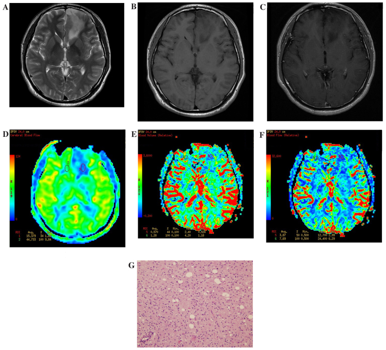
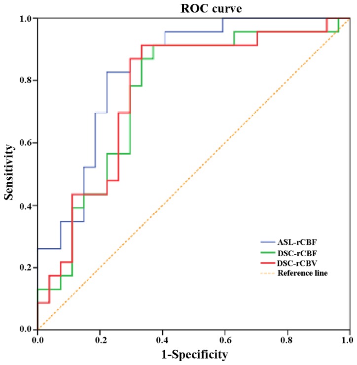
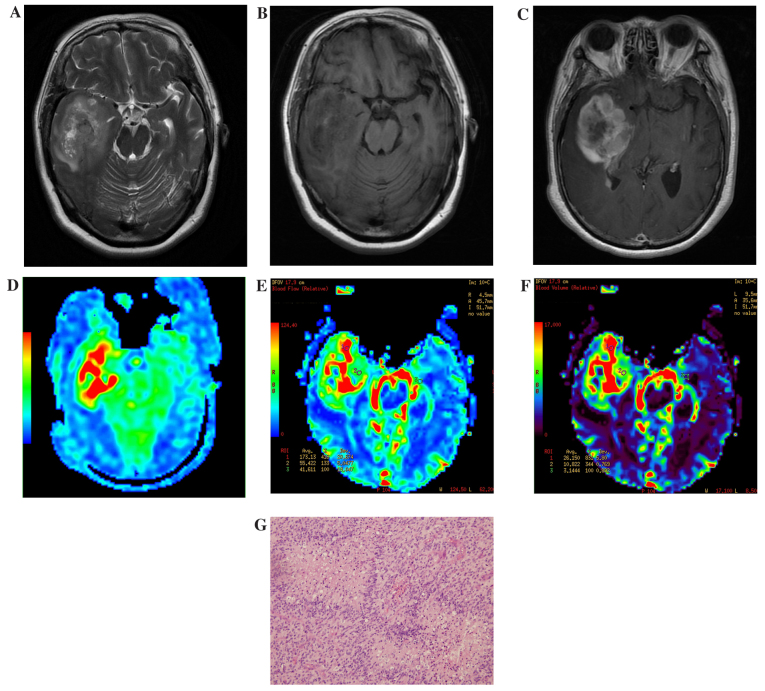
The current study aimed to investigate whole-brain three-dimensional arterial spin labeling imaging (3D ASL) and dynamic susceptibility contrast perfusion-weighted imaging (DSC-PWI), in regards to their diagnostic value of preoperative glioma grade. The parameter values obtained after correction will be correlated with the diagnostic value of 3D ASL and DSC-PWI perfusion. In the current study, 50 patients with gliomas confirmed by pathology were used, including 27 low-grade gliomas (LGGs) and 23 high-grade gliomas (HGGs). Prior to surgery all patients underwent 3 Tesla magnetic resonance imaging (MRI), 3D ASL, DSC-PWI and conventional enhanced MRI scans to obtain original 3D ASL and DSC-PWI images, and the tumor regions with the most obvious parenchyma perfusion and contralateral normal white matter were selected. In these areas, the ASL-relative cerebral blood flow (ASL-rCBF), DSC-relative cerebral blood flow (DSC-rCBF) and DSC-relative cerebral blood volume (DSC-rCBV) parameter values were then obtained after correction for individual differences. The results of the present study show that ASL-CBF, DSC-CBF, DSC-CBV values and ASL-rCBF, DSC-rCBF, DSC-rCBV values increased as the grade of the glioma being imaged increased, and there was a marked difference between the HGGs and the LGGs. ASL-rCBF was significantly positively correlated with DSC-rCBF (r=0.580, P<0.01). In addition, ASL-rCBF was significantly positively correlated with DSC-rCBV (r=0.431, P<0.01). Receiver operating characteristic (ROC) curves were applied to compare the two perfusion parameters of DSC-PWI and 3D ASL in the diagnosis of glioma grade. ASL-rCBF had the highest area value under the ROC curve (0.836). The areas under the ROC curve of DSC-rCBF and DSC-rCBV were analyzed using the Z test, but the difference was not statistically significant. When ASL-rCBF, DSC-rCBF and DSC-rCBV were cutoff at 2.24, 1.85 and 1.68, the sensitivity of HGG diagnosis was 83.2, 91.3 and 91.3%, and the specificity was 77.7, 63.9 and 66.7%, respectively.
本研究旨在探讨全脑三维动脉自旋标记成像(3D ASL)和动态磁敏感对比灌注加权成像(DSC-PWI)对术前胶质瘤分级的诊断价值。校正后获得的参数值将与3D ASL和DSC-PWI灌注的诊断价值相关联。在本研究中,使用了50例经病理证实的胶质瘤患者,包括27例低级别胶质瘤(LGG)和23例高级别胶质瘤(HGG)。手术前,所有患者均接受3特斯拉磁共振成像(MRI)、3D ASL、DSC-PWI和常规增强MRI扫描,以获取原始的3D ASL和DSC-PWI图像,并选择实质灌注最明显的肿瘤区域和对侧正常白质。在这些区域,校正个体差异后获得ASL相对脑血流量(ASL-rCBF)、DSC相对脑血流量(DSC-rCBF)和DSC相对脑血容量(DSC-rCBV)参数值。本研究结果表明,随着成像的胶质瘤分级增加,ASL-CBF、DSC-CBF、DSC-CBV值以及ASL-rCBF、DSC-rCBF、DSC-rCBV值均升高,HGG与LGG之间存在显著差异。ASL-rCBF与DSC-rCBF显著正相关(r=0.580,P<0.01)。此外,ASL-rCBF与DSC-rCBV显著正相关(r=0.431,P<0.01)。应用受试者工作特征(ROC)曲线比较DSC-PWI和3D ASL的两种灌注参数对胶质瘤分级的诊断价值。ASL-rCBF在ROC曲线下的面积值最高(0.836)。使用Z检验分析DSC-rCBF和DSC-rCBV的ROC曲线下面积,但差异无统计学意义。当ASL-rCBF、DSC-rCBF和DSC-rCBV的截断值分别为2.24、1.85和1.68时,HGG诊断的灵敏度分别为83.2%、91.3%和91.3%,特异性分别为77.7%、63.9%和66.7%。