Xu Qian, Liu Qi, Ge Haitao, Ge Xinting, Wu Jiangfen, Qu Jianxun, Xu Kai
The First School of Clinical Medicine, Nanjing Medical University Department of Radiology, Affiliated Hospital of Xuzhou Medical University Department of Medical Imaging, Xuzhou Medical University GE Healthcare, Shanghai, China.
Medicine (Baltimore). 2017 Dec;96(50):e9332. doi: 10.1097/MD.0000000000009332.
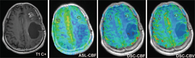
Gliomas constitute over 90% of primary brain tumors. Accurate identification of glioma recurrence and treatment effects is important, as it can help determine whether to continue with standard adjuvant chemotherapy or to switch to a second-line therapy for recurrence. Our purpose is to compare three dimensional pseudo-continuous arterial spin labeling (3D-pcASL) technique and dynamic susceptibility contrast perfusion magnetic resonance imaging (DSC-MRI) for differentiation tumor recurrence from treatment-related effects in gliomas.
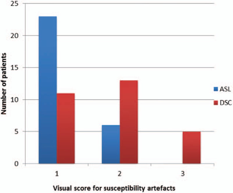
Twenty-nine patients with gliomas previously who showed enlarged, contrast-enhancing lesions within the radiation field after surgery and concurrent chemoradiotherapy (CCRT) were assessed with 3D-pcASL and DSC-MRI. These patients were classified into 2 groups, tumor recurrence group (n = 17) and treatment effects group (n = 12), based on pathologic analysis or clinical-radiologic follow-up. The perfusion imaging quality was assessed using a 3-point scale (1 = poor imaging, 2 = moderate imaging, and 3 = good imaging). Comparison for perfusion imaging-quality score between the 2 techniques was performed with Wilcoxon one-sample test. Quantitative analyses were performed between the 2 groups with cerebral blood flow values (ASL-CBF), relative cerebral blood flow values (ASL-rCBF, DSC-rCBF), and relative cerebral blood volume values (DSC-rCBV) using Wilcoxon one-sample test. The intra-class correlation coefficient (ICC) statistics were calculated for testing intrareader variability in regions of interest (ROIs) measurement of all perfusion parameters.
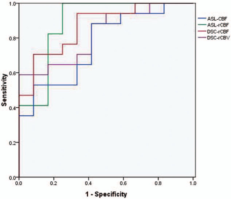
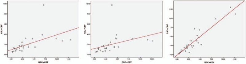
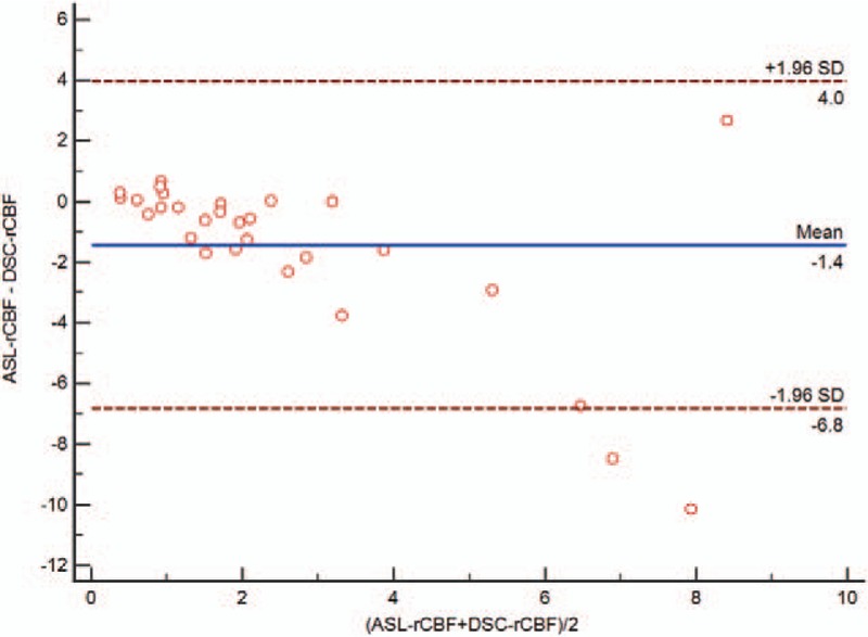
The imaging-quality score of 3D-pcASL was higher than that of DSC-MRI (P = .01). The perfusion parameters between tumor recurrence group and treatment effects group had statistically significant differences. There was a significant correlation between ASL-rCBF and DSC-rCBF values (r = 0.803), between ASL-rCBF and DSC-rCBV values (r = 0.763), and between DSC-rCBF and DSC-rCBV (r = 0.907). A receiver operating characteristic (ROC) curve analysis was performed for significant results of perfusion parameters between the 2 groups. Using a cutoff value of 1.110, ASL-rCBF showed the maximum area under the ROC curve (AUC). However, there were no significant differences among different AUCs. The ICC demonstrated excellent agreement for ROIs measurements of ASL-CBF (ICC = 0.9636), dynamic susceptibility contrast- cerebral blood flow (DSC-CBF) (ICC = 0.8508), and dynamic susceptibility contrast-cerebral blood volume (DSC-CBV) (ICC = 0.8543).
3D-pcASL is an alternative perfusion method to DSC-MRI for the differentiation between tumor recurrence and treatment effects in gliomas. 3D-pcASL is noninvasive and shows fewer susceptibility artifacts than DSC-MRI.
胶质瘤占原发性脑肿瘤的90%以上。准确识别胶质瘤复发和治疗效果很重要,因为这有助于确定是继续进行标准辅助化疗还是转向二线复发治疗。我们的目的是比较三维伪连续动脉自旋标记(3D-pcASL)技术和动态磁敏感对比灌注磁共振成像(DSC-MRI)在区分胶质瘤肿瘤复发与治疗相关效应方面的差异。
对29例先前患有胶质瘤的患者进行评估,这些患者在手术及同步放化疗(CCRT)后,放射野内出现强化增大的病灶,采用3D-pcASL和DSC-MRI检查。根据病理分析或临床-放射学随访,将这些患者分为两组,肿瘤复发组(n = 17)和治疗效应组(n = 12)。灌注成像质量采用3分制评估(1 = 成像差,2 = 成像中等,3 = 成像良好)。采用Wilcoxon单样本检验对两种技术的灌注成像质量评分进行比较。使用Wilcoxon单样本检验对两组间的脑血流量值(ASL-CBF)、相对脑血流量值(ASL-rCBF、DSC-rCBF)和相对脑血容量值(DSC-rCBV)进行定量分析。计算组内相关系数(ICC)统计量,以测试所有灌注参数在感兴趣区域(ROI)测量中的阅片者内变异性。
3D-pcASL的成像质量评分高于DSC-MRI(P = 0.01)。肿瘤复发组和治疗效应组之间的灌注参数有统计学显著差异。ASL-rCBF与DSC-rCBF值之间(r = 0.803)、ASL-rCBF与DSC-rCBV值之间(r = 0.763)以及DSC-rCBF与DSC-rCBV之间(r = 0.907)存在显著相关性。对两组间灌注参数的显著结果进行了受试者工作特征(ROC)曲线分析。使用截断值1.110时,ASL-rCBF在ROC曲线下面积最大(AUC)。然而,不同AUC之间无显著差异。ICC显示,ASL-CBF(ICC = 0.9636)、动态磁敏感对比脑血流量(DSC-CBF)(ICC = 0.8508)和动态磁敏感对比脑血容量(DSC-CBV)(ICC = 0.8543)在ROI测量方面具有良好的一致性。
3D-pcASL是一种替代DSC-MRI的灌注方法,可用于区分胶质瘤的肿瘤复发与治疗效应。3D-pcASL是非侵入性的,与DSC-MRI相比,其磁敏感伪影较少。