Guida Lelio, Stumpo Vittorio, Bellomo Jacopo, van Niftrik Christiaan Hendrik Bas, Sebök Martina, Berhouma Moncef, Bink Andrea, Weller Michael, Kulcsar Zsolt, Regli Luca, Fierstra Jorn
Department of Neurosurgery, University Hospital Zurich, 8091 Zurich, Switzerland.
Clinical Neuroscience Center, University Hospital Zurich, University of Zurich, 8057 Zurich, Switzerland.
Cancers (Basel). 2022 Mar 10;14(6):1432. doi: 10.3390/cancers14061432.
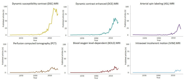
Diffuse gliomas are the most common primary malignant intracranial neoplasms. Aside from the challenges pertaining to their treatment-glioblastomas, in particular, have a dismal prognosis and are currently incurable-their pre-operative assessment using standard neuroimaging has several drawbacks, including broad differentials diagnosis, imprecise characterization of tumor subtype and definition of its infiltration in the surrounding brain parenchyma for accurate resection planning. As the pathophysiological alterations of tumor tissue are tightly linked to an aberrant vascularization, advanced hemodynamic imaging, in addition to other innovative approaches, has attracted considerable interest as a means to improve diffuse glioma characterization. In the present part A of our two-review series, the fundamental concepts, techniques and parameters of hemodynamic imaging are discussed in conjunction with their potential role in the differential diagnosis and grading of diffuse gliomas. In particular, recent evidence on dynamic susceptibility contrast, dynamic contrast-enhanced and arterial spin labeling magnetic resonance imaging are reviewed together with perfusion-computed tomography. While these techniques have provided encouraging results in terms of their sensitivity and specificity, the limitations deriving from a lack of standardized acquisition and processing have prevented their widespread clinical adoption, with current efforts aimed at overcoming the existing barriers.
弥漫性胶质瘤是最常见的原发性颅内恶性肿瘤。除了治疗方面的挑战(尤其是胶质母细胞瘤,预后很差且目前无法治愈)外,使用标准神经影像学进行术前评估存在几个缺点,包括鉴别诊断范围广、肿瘤亚型特征描述不准确以及难以确定其在周围脑实质中的浸润范围以进行精确的切除规划。由于肿瘤组织的病理生理改变与异常血管生成密切相关,除其他创新方法外,先进的血流动力学成像作为改善弥漫性胶质瘤特征描述的一种手段已引起了相当大的关注。在我们两部分综述系列的本部分A中,将结合血流动力学成像在弥漫性胶质瘤鉴别诊断和分级中的潜在作用,讨论其基本概念、技术和参数。特别是,将对动态磁敏感对比、动态对比增强和动脉自旋标记磁共振成像以及灌注计算机断层扫描的最新证据进行综述。虽然这些技术在敏感性和特异性方面取得了令人鼓舞的结果,但由于缺乏标准化的采集和处理方法而产生的局限性阻碍了它们在临床上的广泛应用,目前正在努力克服这些现有障碍。