Dalton Marshall A, Zeidman Peter, Barry Daniel N, Williams Elaine, Maguire Eleanor A
Wellcome Trust Centre for Neuroimaging, Institute of Neurology, University College London, London, UK.
Brain Neurosci Adv. 2017 Apr 6;1:2398212817701448. doi: 10.1177/2398212817701448. eCollection 2017.
The hippocampus plays a central role in cognition, and understanding the specific contributions of its subregions will likely be key to explaining its wide-ranging functions. However, delineating substructures within the human hippocampus in vivo from magnetic resonance image scans is fraught with difficulties. To our knowledge, the extant literature contains only brief descriptions of segmentation procedures used to delineate hippocampal subregions in magnetic resonance imaging/functional magnetic resonance imaging studies.
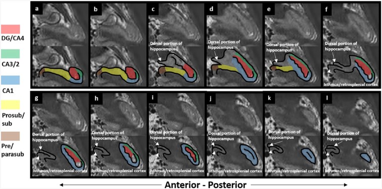
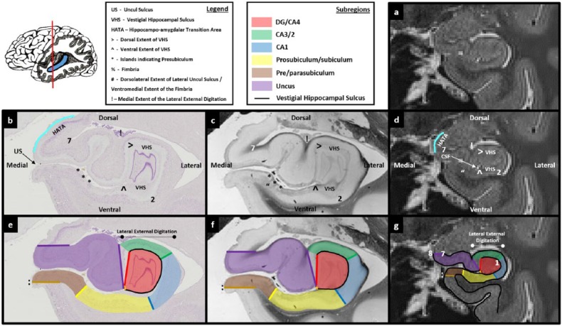
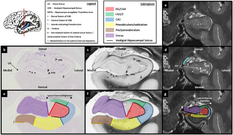
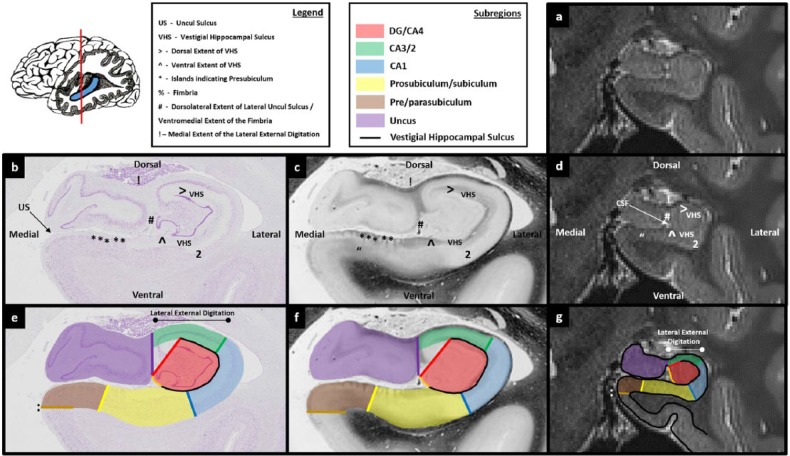
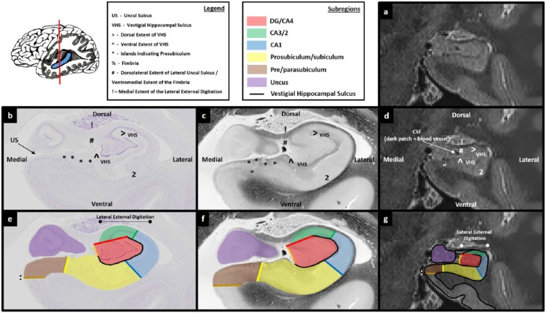
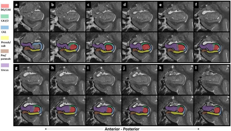
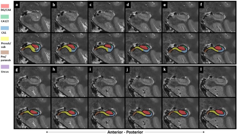
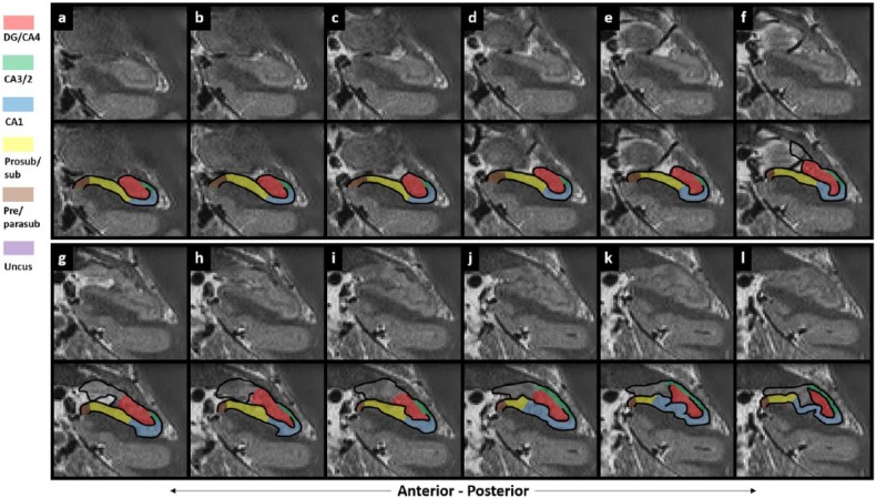
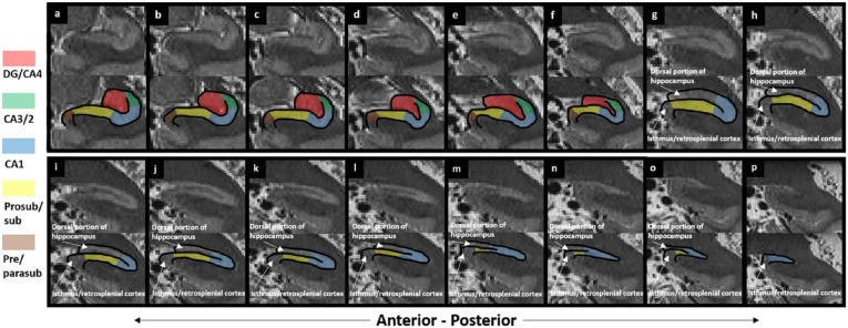
Consequently, here we provide a clear, step-by-step and fully illustrated guide to segmenting hippocampal subregions along the entire length of the human hippocampus on 3T magnetic resonance images.
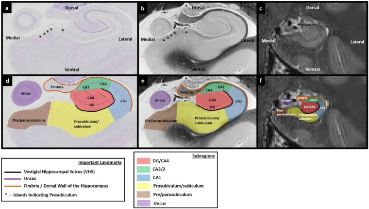
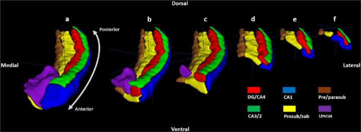
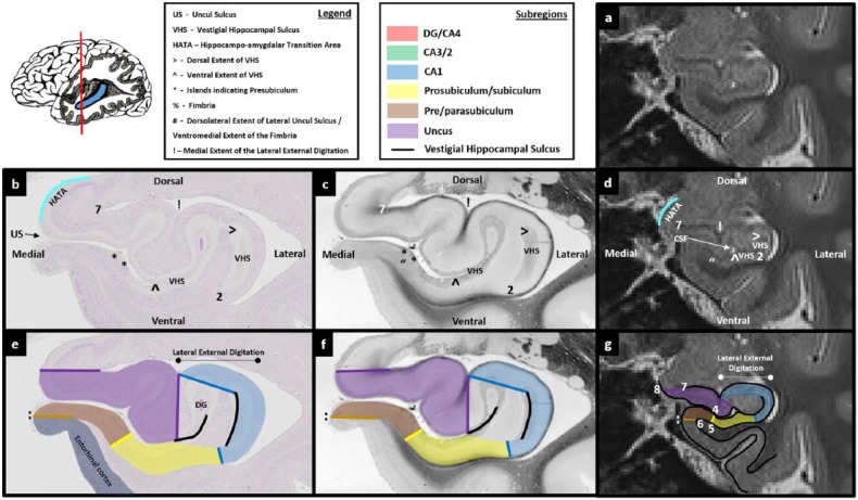
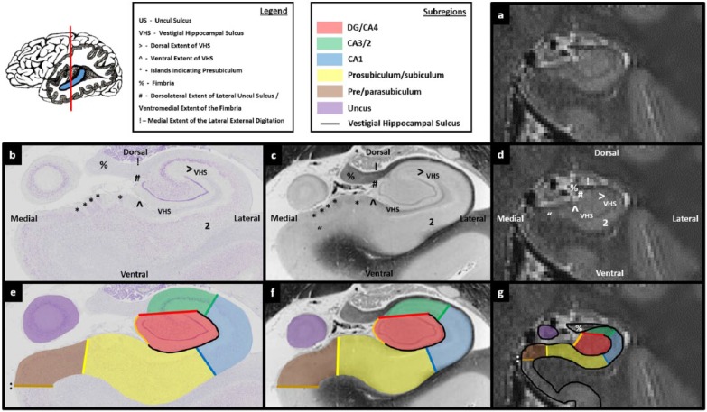
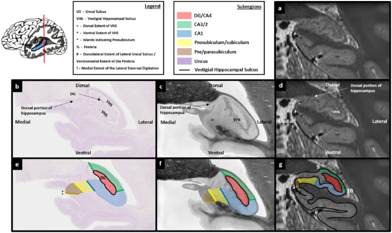
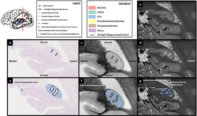
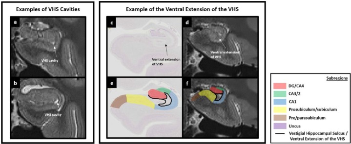
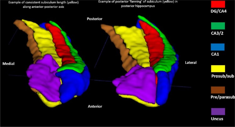
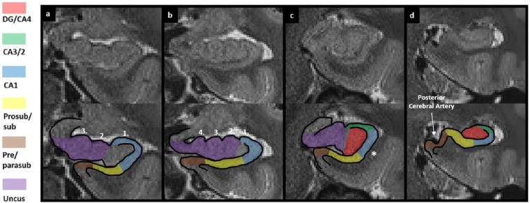
We give a detailed description of how to segment the hippocampus into the following six subregions: dentate gyrus/Cornu Ammonis 4, CA3/2, CA1, subiculum, pre/parasubiculum and the uncus. Importantly, this in-depth protocol incorporates the most recent cyto- and chemo-architectural evidence and includes a series of comprehensive figures which compare slices of histologically stained tissue with equivalent 3T images.
As hippocampal subregion segmentation is an evolving field of research, we do not suggest this protocol is definitive or final. Rather, we present a fully explained and expedient method of manual segmentation which remains faithful to our current understanding of human hippocampal neuroanatomy. We hope that this 'tutorial'-style guide, which can be followed by experts and non-experts alike, will be a practical resource for clinical and research scientists with an interest in the human hippocampus.
海马体在认知中起着核心作用,了解其亚区域的具体贡献可能是解释其广泛功能的关键。然而,通过磁共振图像扫描在体内描绘人类海马体中的亚结构充满困难。据我们所知,现有文献中仅有对磁共振成像/功能磁共振成像研究中用于描绘海马体亚区域的分割程序的简短描述。
因此,我们在此提供一份清晰、逐步且配有详细插图的指南,用于在3T磁共振图像上沿着人类海马体的全长分割海马体亚区域。
我们详细描述了如何将海马体分割为以下六个亚区域:齿状回/海马4区、CA3/2区、CA1区、下托、前/副下托和钩回。重要的是,这一深入的方案纳入了最新的细胞和化学结构证据,并包括一系列将组织学染色组织切片与等效3T图像进行比较的综合图。
由于海马体亚区域分割是一个不断发展的研究领域,我们并不认为该方案是确定的或最终的。相反,我们提出了一种经过充分解释且便捷的手动分割方法,该方法与我们目前对人类海马体神经解剖学的理解一致。我们希望这种“教程”式指南,无论是专家还是非专家都可以遵循,将为对人类海马体感兴趣的临床和研究科学家提供实用资源。