• 文献检索
  • 文档翻译
  • 深度研究
  • 学术资讯
  • Suppr Zotero 插件Zotero 插件
  • 邀请有礼
  • 套餐&价格
  • 历史记录
应用&插件
Suppr Zotero 插件Zotero 插件浏览器插件Mac 客户端Windows 客户端微信小程序
定价
高级版会员购买积分包购买API积分包
服务
文献检索文档翻译深度研究API 文档MCP 服务
关于我们
关于 Suppr公司介绍联系我们用户协议隐私条款
关注我们

Suppr 超能文献

核心技术专利:CN118964589B侵权必究
粤ICP备2023148730 号-1Suppr @ 2026

文献检索

告别复杂PubMed语法,用中文像聊天一样搜索,搜遍4000万医学文献。AI智能推荐,让科研检索更轻松。

立即免费搜索

文件翻译

保留排版,准确专业,支持PDF/Word/PPT等文件格式,支持 12+语言互译。

免费翻译文档

深度研究

AI帮你快速写综述,25分钟生成高质量综述,智能提取关键信息,辅助科研写作。

立即免费体验

骨形态发生蛋白信号通路通过tbx2b和id2a调控斑马鱼中胆管驱动的肝脏再生。

Bone morphogenetic protein signaling governs biliary-driven liver regeneration in zebrafish through tbx2b and id2a.

作者信息

Choi Tae-Young, Khaliq Mehwish, Tsurusaki Shinya, Ninov Nikolay, Stainier Didier Y R, Tanaka Minoru, Shin Donghun

机构信息

Department of Developmental Biology, Pittsburgh Liver Research Center, McGowan Institute for Regenerative Medicine, University of Pittsburgh, Pittsburgh, PA.

Department of Regenerative Medicine, Research Institute, National Center for Global Health and Medicine, Tokyo, Japan.

出版信息

Hepatology. 2017 Nov;66(5):1616-1630. doi: 10.1002/hep.29309. Epub 2017 Sep 29.

DOI:10.1002/hep.29309
PMID:28599080
原文链接:https://pmc.ncbi.nlm.nih.gov/articles/PMC5650528/
Abstract

UNLABELLED

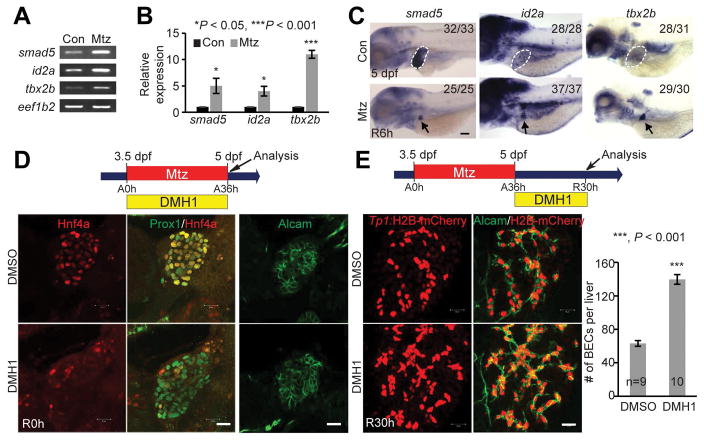
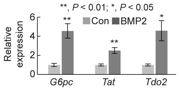
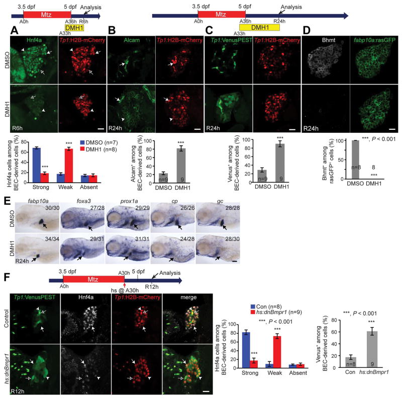
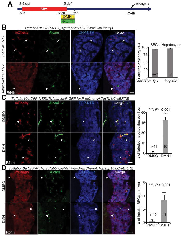
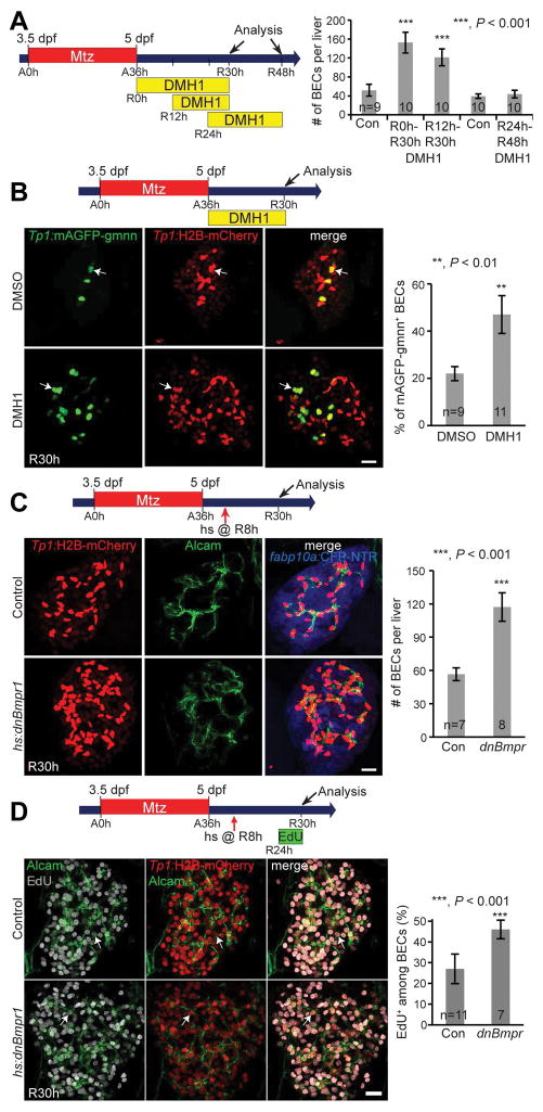
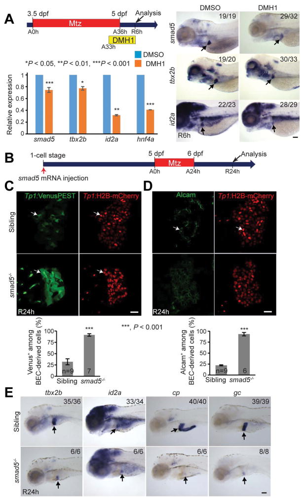
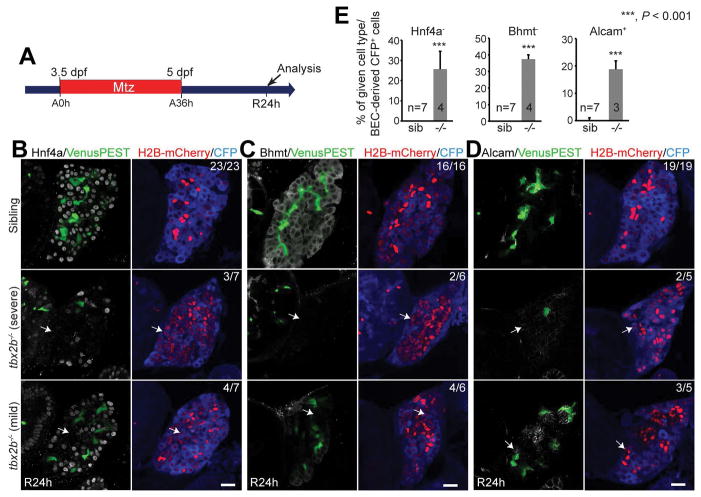
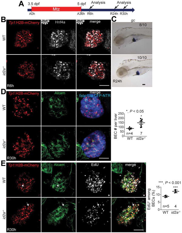
Upon mild liver injury, new hepatocytes originate from preexisting hepatocytes. However, if hepatocyte proliferation is impaired, a manifestation of severe liver injury, biliary epithelial cells (BECs) contribute to new hepatocytes through BEC dedifferentiation into liver progenitor cells (LPCs), also termed oval cells or hepatoblast-like cells (HB-LCs), and subsequent differentiation into hepatocytes. Despite the identification of several factors regulating BEC dedifferentiation and activation, little is known about factors involved in the regulation of LPC differentiation into hepatocytes during liver regeneration. Using a zebrafish model of near-complete hepatocyte ablation, we show that bone morphogenetic protein (Bmp) signaling is required for BEC conversion to hepatocytes, particularly for LPC differentiation into hepatocytes. We found that severe liver injury led to the up-regulation of genes involved in Bmp signaling, including smad5, tbx2b, and id2a, in the liver. Bmp suppression did not block BEC dedifferentiation into HB-LCs; however, the differentiation of HB-LCs into hepatocytes was impaired due to the maintenance of HB-LCs in an undifferentiated state. Later Bmp suppression did not affect HB-LC differentiation but increased BEC number through proliferation. Notably, smad5, tbx2b, and id2a mutants exhibited similar liver regeneration defects as those observed in Bmp-suppressed livers. Moreover, BMP2 addition promoted the differentiation of a murine LPC line into hepatocytes in vitro.

CONCLUSIONS

Bmp signaling regulates BEC-driven liver regeneration through smad5, tbx2b, and id2a: it regulates HB-LC differentiation into hepatocytes through tbx2b and BEC proliferation through id2a; our findings provide insights into promoting innate liver regeneration as a novel therapy. (Hepatology 2017;66:1616-1630).

摘要

未标记

在轻度肝损伤时,新的肝细胞源自已有的肝细胞。然而,如果肝细胞增殖受损,这是严重肝损伤的一种表现,胆管上皮细胞(BECs)会通过BEC去分化为肝祖细胞(LPCs),也称为卵圆细胞或肝母样细胞(HB-LCs),随后分化为肝细胞,从而对新肝细胞的形成做出贡献。尽管已经鉴定出了几种调节BEC去分化和激活的因子,但对于肝再生过程中LPC分化为肝细胞的调节因子知之甚少。利用斑马鱼近乎完全肝细胞消融的模型,我们表明骨形态发生蛋白(Bmp)信号通路对于BEC转化为肝细胞是必需的,特别是对于LPC分化为肝细胞。我们发现严重肝损伤导致肝脏中参与Bmp信号通路的基因上调,包括smad5、tbx2b和id2a。Bmp抑制并未阻止BEC去分化为HB-LCs;然而,由于HB-LCs维持在未分化状态,HB-LCs向肝细胞的分化受到损害。后期Bmp抑制并不影响HB-LC分化,但通过增殖增加了BEC数量。值得注意的是,smad5、tbx2b和id2a突变体表现出与Bmp抑制肝脏中观察到的类似的肝再生缺陷。此外,添加BMP2可促进小鼠LPC系在体外分化为肝细胞。

结论

Bmp信号通路通过smad5、tbx2b和id2a调节BEC驱动的肝再生:它通过tbx2b调节HB-LC分化为肝细胞,并通过id2a调节BEC增殖;我们的发现为促进先天性肝再生作为一种新疗法提供了见解。(《肝脏病学》2017年;66:1616 - 1630)

https://cdn.ncbi.nlm.nih.gov/pmc/blobs/ac0e/5650528/86a558687cf3/nihms883515f8.jpg
https://cdn.ncbi.nlm.nih.gov/pmc/blobs/ac0e/5650528/a447804d6c1b/nihms883515f1.jpg
https://cdn.ncbi.nlm.nih.gov/pmc/blobs/ac0e/5650528/da22f8c78ae8/nihms883515f2.jpg
https://cdn.ncbi.nlm.nih.gov/pmc/blobs/ac0e/5650528/117ddc43de9b/nihms883515f3.jpg
https://cdn.ncbi.nlm.nih.gov/pmc/blobs/ac0e/5650528/e60a8ef1863a/nihms883515f4.jpg
https://cdn.ncbi.nlm.nih.gov/pmc/blobs/ac0e/5650528/19e1e87ea1b5/nihms883515f5.jpg
https://cdn.ncbi.nlm.nih.gov/pmc/blobs/ac0e/5650528/6621dbcf151a/nihms883515f6.jpg
https://cdn.ncbi.nlm.nih.gov/pmc/blobs/ac0e/5650528/2561a54b96ff/nihms883515f7.jpg
https://cdn.ncbi.nlm.nih.gov/pmc/blobs/ac0e/5650528/86a558687cf3/nihms883515f8.jpg

相似文献

1
Bone morphogenetic protein signaling governs biliary-driven liver regeneration in zebrafish through tbx2b and id2a.骨形态发生蛋白信号通路通过tbx2b和id2a调控斑马鱼中胆管驱动的肝脏再生。
Hepatology. 2017 Nov;66(5):1616-1630. doi: 10.1002/hep.29309. Epub 2017 Sep 29.
2
Bromodomain and extraterminal (BET) proteins regulate biliary-driven liver regeneration.溴结构域与额外末端(BET)蛋白调控胆管驱动的肝脏再生。
J Hepatol. 2016 Feb;64(2):316-325. doi: 10.1016/j.jhep.2015.10.017. Epub 2015 Oct 24.
3
Farnesoid X Receptor Activation Impairs Liver Progenitor Cell-Mediated Liver Regeneration via the PTEN-PI3K-AKT-mTOR Axis in Zebrafish.法尼醇 X 受体激活通过 PTEN-PI3K-AKT-mTOR 轴损害肝祖细胞介导的斑马鱼肝脏再生。
Hepatology. 2021 Jul;74(1):397-410. doi: 10.1002/hep.31679. Epub 2021 Jun 15.
4
Hdac1 Regulates Differentiation of Bipotent Liver Progenitor Cells During Regeneration via Sox9b and Cdk8.Hdac1 通过 Sox9b 和 Cdk8 调控肝祖细胞的双向分化在再生过程中。
Gastroenterology. 2019 Jan;156(1):187-202.e14. doi: 10.1053/j.gastro.2018.09.039. Epub 2018 Sep 26.
5
Stat3 Regulates Liver Progenitor Cell-Driven Liver Regeneration in Zebrafish.Stat3调控斑马鱼中肝祖细胞驱动的肝脏再生
Gene Expr. 2018 Aug 22;18(3):157-170. doi: 10.3727/105221618X15242506133273. Epub 2018 Apr 24.
6
Mammalian Target of Rapamycin Complex 1 Signaling Is Required for the Dedifferentiation From Biliary Cell to Bipotential Progenitor Cell in Zebrafish Liver Regeneration.哺乳动物雷帕霉素靶蛋白复合物 1 信号通路在斑马鱼肝脏再生中胆管细胞向双潜能祖细胞去分化过程中发挥作用。
Hepatology. 2019 Dec;70(6):2092-2106. doi: 10.1002/hep.30790. Epub 2019 Aug 12.
7
Knockdown-Mediated Hepatocyte Damages Elicit Both the Formation of Hybrid Hepatocytes and Biliary Conversion to Hepatocytes in Zebrafish Larvae.敲低介导的肝细胞损伤引发斑马鱼幼虫中杂交肝细胞的形成以及胆管向肝细胞的转化。
Gene Expr. 2017 Jul 7;17(3):237-249. doi: 10.3727/105221617X695195. Epub 2017 Mar 1.
8
Id2a is required for hepatic outgrowth during liver development in zebrafish.Id2a在斑马鱼肝脏发育过程中的肝脏生长中是必需的。
Mech Dev. 2015 Nov;138 Pt 3:399-414. doi: 10.1016/j.mod.2015.05.001. Epub 2015 May 27.
9
Extensive conversion of hepatic biliary epithelial cells to hepatocytes after near total loss of hepatocytes in zebrafish.在斑马鱼中,近全量肝细胞丢失后,肝脏胆管上皮细胞广泛转化为肝细胞。
Gastroenterology. 2014 Mar;146(3):776-88. doi: 10.1053/j.gastro.2013.10.019. Epub 2013 Oct 19.
10
Notch Inhibition Promotes Differentiation of Liver Progenitor Cells into Hepatocytes via Repression in Zebrafish.Notch抑制通过在斑马鱼中的抑制作用促进肝祖细胞向肝细胞分化。
Stem Cells Int. 2019 Mar 12;2019:8451282. doi: 10.1155/2019/8451282. eCollection 2019.

引用本文的文献

1
Triggering Mechanisms of Hepatocyte Repopulation during Liver Regeneration.肝脏再生过程中肝细胞再填充的触发机制
Biomol Ther (Seoul). 2025 Jul 1;33(4):582-593. doi: 10.4062/biomolther.2025.035. Epub 2025 Jun 12.
2
Cholangiocytes contribute to hepatocyte regeneration after partial liver injury during growth spurt in zebrafish.在斑马鱼生长突增期间,胆管细胞在部分肝损伤后对肝细胞再生有贡献。
Nat Commun. 2025 Jun 6;16(1):5260. doi: 10.1038/s41467-025-60334-y.
3
Hsa-miR-637 inhibits human hepatocyte proliferation by targeting Med1-interacting proteins.

本文引用的文献

1
Spatially Resolved Genome-wide Transcriptional Profiling Identifies BMP Signaling as Essential Regulator of Zebrafish Cardiomyocyte Regeneration.空间分辨全基因组转录谱分析鉴定 BMP 信号作为斑马鱼心肌细胞再生的必要调节因子。
Dev Cell. 2016 Jan 11;36(1):36-49. doi: 10.1016/j.devcel.2015.12.010. Epub 2015 Dec 31.
2
Bromodomain and extraterminal (BET) proteins regulate biliary-driven liver regeneration.溴结构域与额外末端(BET)蛋白调控胆管驱动的肝脏再生。
J Hepatol. 2016 Feb;64(2):316-325. doi: 10.1016/j.jhep.2015.10.017. Epub 2015 Oct 24.
3
Hepatic progenitor cells of biliary origin with liver repopulation capacity.
人源微小RNA-637通过靶向中介蛋白1相互作用蛋白抑制人肝细胞增殖。
Liver Res. 2021 Jan 23;5(2):88-96. doi: 10.1016/j.livres.2021.01.002. eCollection 2021 Jun.
4
Molecular mechanisms in liver repair and regeneration: from physiology to therapeutics.肝脏修复与再生的分子机制:从生理学到治疗学
Signal Transduct Target Ther. 2025 Feb 8;10(1):63. doi: 10.1038/s41392-024-02104-8.
5
Using different zebrafish models to explore liver regeneration.使用不同的斑马鱼模型探索肝脏再生。
Front Cell Dev Biol. 2024 Oct 31;12:1485773. doi: 10.3389/fcell.2024.1485773. eCollection 2024.
6
Deletion of Tgm2 suppresses BMP-mediated hepatocyte-to-cholangiocyte metaplasia in ductular reaction.Tgm2 的缺失抑制了 BMP 介导的胆管细胞化生在胆管反应中的作用。
Cell Prolif. 2024 Oct;57(10):e13646. doi: 10.1111/cpr.13646. Epub 2024 Apr 16.
7
Determining Photoreceptor Cell Identity: Rod Versus Cone Fate Governed by tbx2b Opposing nrl.确定光感受器细胞身份:tbx2b 拮抗 nrl 决定杆状细胞与锥状细胞命运。
Invest Ophthalmol Vis Sci. 2024 Jan 2;65(1):39. doi: 10.1167/iovs.65.1.39.
8
Signaling pathways of liver regeneration: Biological mechanisms and implications.肝脏再生的信号通路:生物学机制及意义
iScience. 2023 Dec 7;27(1):108683. doi: 10.1016/j.isci.2023.108683. eCollection 2024 Jan 19.
9
PPARα activation promotes liver progenitor cell-mediated liver regeneration by suppressing YAP signaling in zebrafish.过氧化物酶体增殖物激活受体 α 的激活通过抑制斑马鱼中 YAP 信号通路促进肝祖细胞介导的肝再生。
Sci Rep. 2023 Oct 25;13(1):18312. doi: 10.1038/s41598-023-44935-5.
10
Arouse potential stemness: Intrinsic and acquired stem cell therapeutic strategies for advanced liver diseases.激发潜在干性:晚期肝病的内在和获得性干细胞治疗策略。
Cell Insight. 2023 Aug 11;2(5):100115. doi: 10.1016/j.cellin.2023.100115. eCollection 2023 Oct.
具有肝脏再填充能力的胆管源性肝祖细胞。
Nat Cell Biol. 2015 Aug;17(8):971-983. doi: 10.1038/ncb3203. Epub 2015 Jul 20.
4
Genetic compensation induced by deleterious mutations but not gene knockdowns.有害突变而非基因敲低诱导的遗传补偿。
Nature. 2015 Aug 13;524(7564):230-3. doi: 10.1038/nature14580. Epub 2015 Jul 13.
5
Hepatocyte-specific ablation in zebrafish to study biliary-driven liver regeneration.在斑马鱼中进行肝细胞特异性消融以研究胆管驱动的肝脏再生。
J Vis Exp. 2015 May 20(99):e52785. doi: 10.3791/52785.
6
Id2a is required for hepatic outgrowth during liver development in zebrafish.Id2a在斑马鱼肝脏发育过程中的肝脏生长中是必需的。
Mech Dev. 2015 Nov;138 Pt 3:399-414. doi: 10.1016/j.mod.2015.05.001. Epub 2015 May 27.
7
Control of hepatocyte proliferation and survival by Fgf receptors is essential for liver regeneration in mice.成纤维细胞生长因子受体对肝实质细胞增殖和存活的调控对于小鼠肝脏再生是必需的。
Gut. 2015 Sep;64(9):1444-53. doi: 10.1136/gutjnl-2014-307874.
8
Antagonistic interaction between Wnt and Notch activity modulates the regenerative capacity of a zebrafish fibrotic liver model.Wnt与Notch活性之间的拮抗相互作用调节斑马鱼纤维化肝脏模型的再生能力。
Hepatology. 2014 Nov;60(5):1753-66. doi: 10.1002/hep.27285. Epub 2014 Sep 10.
9
Stem/progenitor cells in liver development, homeostasis, regeneration, and reprogramming.肝脏发育、稳态、再生和重编程中的干细胞/祖细胞。
Cell Stem Cell. 2014 May 1;14(5):561-74. doi: 10.1016/j.stem.2014.04.010.
10
Regeneration of liver after extreme hepatocyte loss occurs mainly via biliary transdifferentiation in zebrafish.在斑马鱼中,极端肝细胞损失后的肝脏再生主要通过胆管细胞转分化发生。
Gastroenterology. 2014 Mar;146(3):789-800.e8. doi: 10.1053/j.gastro.2013.11.045. Epub 2013 Dec 4.