• 文献检索
  • 文档翻译
  • 深度研究
  • 学术资讯
  • Suppr Zotero 插件Zotero 插件
  • 邀请有礼
  • 套餐&价格
  • 历史记录
应用&插件
Suppr Zotero 插件Zotero 插件浏览器插件Mac 客户端Windows 客户端微信小程序
定价
高级版会员购买积分包购买API积分包
服务
文献检索文档翻译深度研究API 文档MCP 服务
关于我们
关于 Suppr公司介绍联系我们用户协议隐私条款
关注我们

Suppr 超能文献

核心技术专利:CN118964589B侵权必究
粤ICP备2023148730 号-1Suppr @ 2026

文献检索

告别复杂PubMed语法,用中文像聊天一样搜索,搜遍4000万医学文献。AI智能推荐,让科研检索更轻松。

立即免费搜索

文件翻译

保留排版,准确专业,支持PDF/Word/PPT等文件格式,支持 12+语言互译。

免费翻译文档

深度研究

AI帮你快速写综述,25分钟生成高质量综述,智能提取关键信息,辅助科研写作。

立即免费体验

攻克淀粉样蛋白:组织蛋白酶L降解α-突触核蛋白的机制洞察

Taking a Bite Out of Amyloid: Mechanistic Insights into α-Synuclein Degradation by Cathepsin L.

作者信息

McGlinchey Ryan P, Dominah Gifty A, Lee Jennifer C

机构信息

Laboratory of Protein Conformation and Dynamics, Biochemistry and Biophysics Center, National Heart, Lung, and Blood Institute, National Institutes of Health , Bethesda, Maryland 20892, United States.

出版信息

Biochemistry. 2017 Aug 1;56(30):3881-3884. doi: 10.1021/acs.biochem.7b00360. Epub 2017 Jun 28.

DOI:10.1021/acs.biochem.7b00360
PMID:28614652
原文链接:https://pmc.ncbi.nlm.nih.gov/articles/PMC5547998/
Abstract

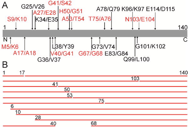
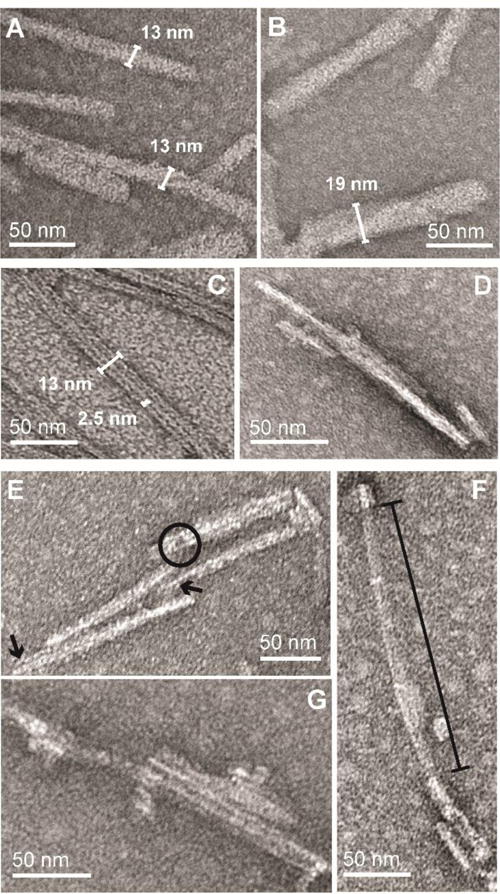
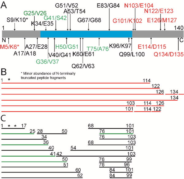
A common hallmark of amyloids is their resistance to an array of proteases, highlighting the difficulty in degrading these disease-related aggregated proteinaceous materials. Here, we report on the potent activity of cathepsin L (CtsL), a lysosomal protease that proteolyzes the Parkinson's disease-related amyloid formed by α-synuclein (α-syn). Using liquid chromatography with mass spectrometry and transmission electron microscopy, an elegant mechanism is revealed on the residue and ultrastructural level, respectively. Specifically, CtsL always truncates α-syn fibrils first at the C-terminus before attacking the internal β-sheet-rich region between residues 30 and 100. This suggests that only upon removal of the α-syn C-terminus can CtsL gain access to residues within the amyloid core. Interestingly, three of the four mapped sites contain a glycine residue (G36, G41, and G51) that is likely to be involved in a β-turn in the fibril, whereupon cutting would lead to solvent exposure of internal residues and allow further proteolysis. Via close inspection of the fibril morphology, products resulting from CtsL degradation show imperfections along the fibril axis, with missing protein density as though they have been cannibalized. The ability of CtsL to degrade α-syn amyloid fibrils offers a promising strategy for improving the cellular clearance of aggregated α-syn through the modulation of protease levels and activity.

摘要

淀粉样蛋白的一个常见特征是它们对一系列蛋白酶具有抗性,这突出了降解这些与疾病相关的聚集蛋白质材料的困难。在此,我们报道了组织蛋白酶L(CtsL)的强大活性,它是一种溶酶体蛋白酶,可蛋白水解由α-突触核蛋白(α-syn)形成的帕金森病相关淀粉样蛋白。分别使用液相色谱-质谱联用和透射电子显微镜,在残基和超微结构水平上揭示了一个精妙的机制。具体而言,CtsL总是先在α-syn原纤维的C末端进行切割,然后再攻击30至100位残基之间富含β-折叠的内部区域。这表明只有在去除α-syn的C末端后,CtsL才能进入淀粉样核心内的残基。有趣的是,四个定位位点中的三个含有一个甘氨酸残基(G36、G41和G51),该残基可能参与原纤维中的β-转角,切割后会导致内部残基暴露于溶剂中并允许进一步的蛋白水解。通过仔细观察原纤维形态,CtsL降解产生的产物沿原纤维轴显示出缺陷,蛋白质密度缺失,就好像它们被自噬了一样。CtsL降解α-syn淀粉样原纤维的能力为通过调节蛋白酶水平和活性来改善聚集α-syn的细胞清除提供了一个有前景的策略。

https://cdn.ncbi.nlm.nih.gov/pmc/blobs/09f4/5547998/f99ca35de3db/nihms888375f3.jpg
https://cdn.ncbi.nlm.nih.gov/pmc/blobs/09f4/5547998/643091116206/nihms888375f1.jpg
https://cdn.ncbi.nlm.nih.gov/pmc/blobs/09f4/5547998/9ee51d220f0c/nihms888375f2.jpg
https://cdn.ncbi.nlm.nih.gov/pmc/blobs/09f4/5547998/f99ca35de3db/nihms888375f3.jpg
https://cdn.ncbi.nlm.nih.gov/pmc/blobs/09f4/5547998/643091116206/nihms888375f1.jpg
https://cdn.ncbi.nlm.nih.gov/pmc/blobs/09f4/5547998/9ee51d220f0c/nihms888375f2.jpg
https://cdn.ncbi.nlm.nih.gov/pmc/blobs/09f4/5547998/f99ca35de3db/nihms888375f3.jpg

相似文献

1
Taking a Bite Out of Amyloid: Mechanistic Insights into α-Synuclein Degradation by Cathepsin L.攻克淀粉样蛋白:组织蛋白酶L降解α-突触核蛋白的机制洞察
Biochemistry. 2017 Aug 1;56(30):3881-3884. doi: 10.1021/acs.biochem.7b00360. Epub 2017 Jun 28.
2
Cathepsin K is a potent disaggregase of α-synuclein fibrils.组织蛋白酶 K 是一种有效的α-突触核蛋白原纤维解聚酶。
Biochem Biophys Res Commun. 2020 Sep 3;529(4):1106-1111. doi: 10.1016/j.bbrc.2020.06.155. Epub 2020 Jul 31.
3
Cysteine cathepsins are essential in lysosomal degradation of α-synuclein.半胱氨酸组织蛋白酶在α-突触核蛋白的溶酶体降解过程中至关重要。
Proc Natl Acad Sci U S A. 2015 Jul 28;112(30):9322-7. doi: 10.1073/pnas.1500937112. Epub 2015 Jul 13.
4
C-terminal α-synuclein truncations are linked to cysteine cathepsin activity in Parkinson's disease.C 端α-突触核蛋白截断与帕金森病中半胱氨酸组织蛋白酶活性有关。
J Biol Chem. 2019 Jun 21;294(25):9973-9984. doi: 10.1074/jbc.RA119.008930. Epub 2019 May 15.
5
Cathepsin L prevents the accumulation of alpha-synuclein fibrils in the cell.组织蛋白酶 L 可防止α-突触核蛋白原纤维在细胞内堆积。
Genes Cells. 2024 Apr;29(4):328-336. doi: 10.1111/gtc.13099. Epub 2024 Feb 17.
6
High-Pressure-Driven Reversible Dissociation of α-Synuclein Fibrils Reveals Structural Hierarchy.高压驱动的α-突触核蛋白原纤维可逆解离揭示了结构层次。
Biophys J. 2017 Oct 17;113(8):1685-1696. doi: 10.1016/j.bpj.2017.08.042.
7
Mechanistic basis for receptor-mediated pathological α-synuclein fibril cell-to-cell transmission in Parkinson's disease.帕金森病中受体介导的病理性α-突触核蛋白纤维细胞间传递的机制基础。
Proc Natl Acad Sci U S A. 2021 Jun 29;118(26). doi: 10.1073/pnas.2011196118.
8
The N terminus of α-synuclein dictates fibril formation.α-突触核蛋白的 N 端决定纤维形成。
Proc Natl Acad Sci U S A. 2021 Aug 31;118(35). doi: 10.1073/pnas.2023487118.
9
The chaperone activity of α-synuclein: Utilizing deletion mutants to map its interaction with target proteins.α-突触核蛋白的伴侣活性:利用缺失突变体来绘制其与靶蛋白的相互作用图谱。
Proteins. 2012 May;80(5):1316-25. doi: 10.1002/prot.24028. Epub 2012 Feb 10.
10
Structural Insights into α-Synuclein Fibril Polymorphism: Effects of Parkinson's Disease-Related C-Terminal Truncations.α-突触核蛋白纤维多态性的结构见解:帕金森病相关 C 末端截断的影响。
J Mol Biol. 2019 Sep 6;431(19):3913-3919. doi: 10.1016/j.jmb.2019.07.001. Epub 2019 Jul 8.

引用本文的文献

1
Recombinant cathepsins B and L promote α-synuclein clearance and restore lysosomal function in human and murine models with α-synuclein pathology.重组组织蛋白酶B和L可促进α-突触核蛋白的清除,并在患有α-突触核蛋白病理学的人类和小鼠模型中恢复溶酶体功能。
Mol Neurodegener. 2025 Aug 29;20(1):95. doi: 10.1186/s13024-025-00886-1.
2
Defining essential charged residues in fibril formation of a lysosomal derived N-terminal α-synuclein truncation.确定溶酶体衍生的N端α-突触核蛋白截短体纤维形成中的必需带电残基。
Nat Commun. 2025 Apr 23;16(1):3825. doi: 10.1038/s41467-025-58899-9.
3
Reciprocal effects of alpha-synuclein aggregation and lysosomal homeostasis in synucleinopathy models.

本文引用的文献

1
Solid-state NMR structure of a pathogenic fibril of full-length human α-synuclein.全长人α-突触核蛋白致病原纤维的固态核磁共振结构
Nat Struct Mol Biol. 2016 May;23(5):409-15. doi: 10.1038/nsmb.3194. Epub 2016 Mar 28.
2
A critical appraisal of the pathogenic protein spread hypothesis of neurodegeneration.神经退行性变的致病蛋白传播假说的批判性评估。
Nat Rev Neurosci. 2016 Apr;17(4):251-60. doi: 10.1038/nrn.2016.13.
3
α-Synuclein-induced lysosomal dysfunction occurs through disruptions in protein trafficking in human midbrain synucleinopathy models.
α-突触核蛋白聚集和溶酶体动态平衡在突触核蛋白病模型中的相互影响。
Transl Neurodegener. 2023 Jun 13;12(1):31. doi: 10.1186/s40035-023-00363-z.
4
Cellular processing of α-synuclein fibrils results in distinct physiological C-terminal truncations with a major cleavage site at residue Glu 114.α-突触核蛋白纤维的细胞加工导致具有独特生理 C 末端截断的明显差异,其主要切割位点位于残基Glu114。
J Biol Chem. 2023 Jul;299(7):104912. doi: 10.1016/j.jbc.2023.104912. Epub 2023 Jun 10.
5
Cathepsin L-containing exosomes from α-synuclein-activated microglia induce neurotoxicity through the P2X7 receptor.来自α-突触核蛋白激活的小胶质细胞的含组织蛋白酶L的外泌体通过P2X7受体诱导神经毒性。
NPJ Parkinsons Dis. 2022 Oct 6;8(1):127. doi: 10.1038/s41531-022-00394-9.
6
The Role of Extracellular Matrix Components in the Spreading of Pathological Protein Aggregates.细胞外基质成分在病理性蛋白质聚集体扩散中的作用
Front Cell Neurosci. 2022 Apr 29;16:844211. doi: 10.3389/fncel.2022.844211. eCollection 2022.
7
Proteolysis of Amyloid β by Lysosomal Enzymes as a Function of Fibril Morphology.溶酶体酶对淀粉样β蛋白的蛋白水解作用与纤维形态的关系
ACS Omega. 2021 Nov 18;6(47):31520-31527. doi: 10.1021/acsomega.1c03915. eCollection 2021 Nov 30.
8
Targeted proteolytic products of τ and α-synuclein in neurodegeneration.神经退行性疾病中τ和α-突触核蛋白的靶向蛋白水解产物。
Essays Biochem. 2021 Dec 22;65(7):905-912. doi: 10.1042/EBC20210028.
9
Current Evidence for a Bidirectional Loop Between the Lysosome and Alpha-Synuclein Proteoforms.溶酶体与α-突触核蛋白蛋白变体之间双向循环的当前证据
Front Cell Dev Biol. 2020 Nov 17;8:598446. doi: 10.3389/fcell.2020.598446. eCollection 2020.
10
Cathepsin K is a potent disaggregase of α-synuclein fibrils.组织蛋白酶 K 是一种有效的α-突触核蛋白原纤维解聚酶。
Biochem Biophys Res Commun. 2020 Sep 3;529(4):1106-1111. doi: 10.1016/j.bbrc.2020.06.155. Epub 2020 Jul 31.
在人类中脑突触核蛋白病模型中,α-突触核蛋白诱导的溶酶体功能障碍是通过蛋白质运输的破坏而发生的。
Proc Natl Acad Sci U S A. 2016 Feb 16;113(7):1931-6. doi: 10.1073/pnas.1520335113. Epub 2016 Feb 2.
4
α-Synuclein Amyloid Fibrils with Two Entwined, Asymmetrically Associated Protofibrils.具有两条缠绕且不对称关联原纤维的α-突触核蛋白淀粉样纤维。
J Biol Chem. 2016 Jan 29;291(5):2310-8. doi: 10.1074/jbc.M115.698787. Epub 2015 Dec 7.
5
Determinants of amyloid fibril degradation by the PDZ protease HTRA1.PDZ 蛋白酶 HTRA1 降解淀粉样纤维的决定因素。
Nat Chem Biol. 2015 Nov;11(11):862-9. doi: 10.1038/nchembio.1931. Epub 2015 Oct 5.
6
Structure of the toxic core of α-synuclein from invisible crystals.来自不可见晶体的α-突触核蛋白毒性核心结构。
Nature. 2015 Sep 24;525(7570):486-90. doi: 10.1038/nature15368. Epub 2015 Sep 9.
7
Cysteine cathepsins are essential in lysosomal degradation of α-synuclein.半胱氨酸组织蛋白酶在α-突触核蛋白的溶酶体降解过程中至关重要。
Proc Natl Acad Sci U S A. 2015 Jul 28;112(30):9322-7. doi: 10.1073/pnas.1500937112. Epub 2015 Jul 13.
8
Amyloid polymorphism: structural basis and neurobiological relevance.淀粉样蛋白多态性:结构基础与神经生物学关联
Neuron. 2015 May 6;86(3):632-45. doi: 10.1016/j.neuron.2015.03.017.
9
The structure of fibrils from 'misfolded' proteins.来自“错误折叠”蛋白质的原纤维结构。
Curr Opin Struct Biol. 2015 Feb;30:43-49. doi: 10.1016/j.sbi.2014.12.001. Epub 2014 Dec 24.
10
Cell-to-cell transmission of pathogenic proteins in neurodegenerative diseases.神经退行性疾病中致病性蛋白的细胞间传递。
Nat Med. 2014 Feb;20(2):130-8. doi: 10.1038/nm.3457.