Department of Geriatrics, Tianjin Medical University General Hospital, Tianjin Geriatrics Institute, Anshan Road No. 154, Tianjin, 300052, China.
Laboratory of Neuro-Trauma and Neurodegenerative Disorder, Tianjin Geriatrics Institute, Tianjin, 300052, China.
Neurochem Res. 2017 Oct;42(10):2892-2901. doi: 10.1007/s11064-017-2310-0. Epub 2017 Jun 15.
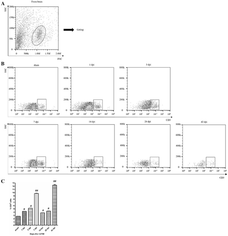
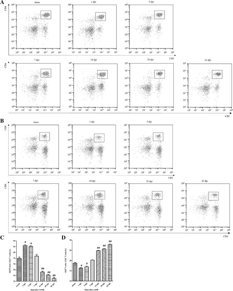
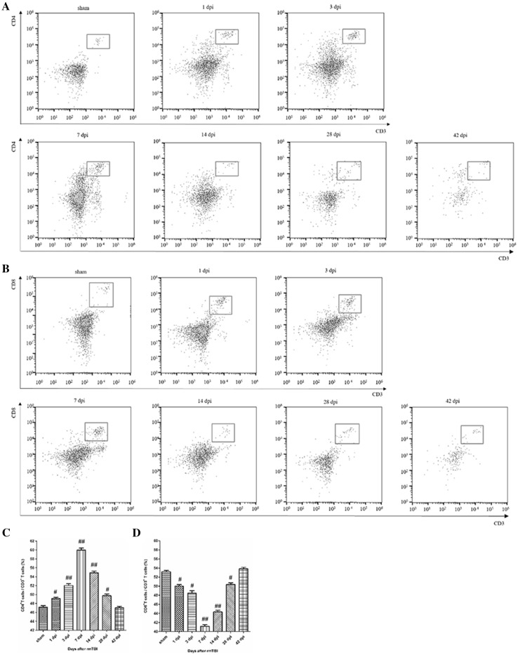
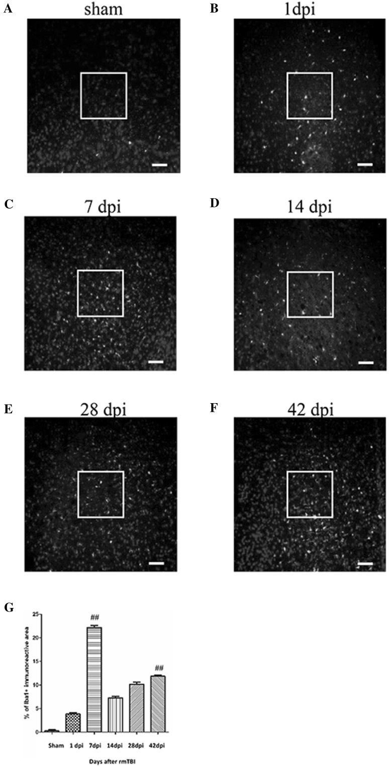
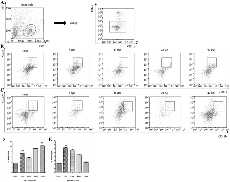
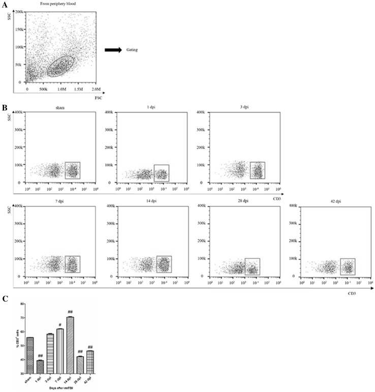
Although, there is growing awareness in the progressive neurodegeneration of chronic traumatic encephalopathy, changes of immune reactions remain equivocal at best. Thus, in a clinically relevant rat repetitive mild traumatic brain injury (rmTBI) model, some immunologic cells (T cell subsets, microglia) in the injured brain and peripheral blood were analyzed by flow cytometry and immunofluorescence. In the injured brain, CD3 T cells showed a bimodal increase during 42 days post-injury (dpi). CD3CD4 T cells firstly increased and then decreased, while CD3CD8 T cells had reversed tendency. CD86/CD11b M1-like microglia increased at 42 dpi and CD206/CD11b M2-like microglia peaked at 7 dpi. In addition, peripheral immune suppression was implicated in the chronic phase after rmTBI. Taken together, the study provided useful information on long-term dynamic changes of some immune cells after rmTBI in rats.
尽管人们越来越意识到慢性创伤性脑病的进行性神经退行性变,但免疫反应的变化充其量也只是模棱两可。因此,在一个具有临床相关性的大鼠重复轻度创伤性脑损伤(rmTBI)模型中,通过流式细胞术和免疫荧光法分析了损伤大脑和外周血中的一些免疫细胞(T 细胞亚群、小胶质细胞)。在损伤的大脑中,CD3 T 细胞在损伤后 42 天(dpi)呈现双峰增加。CD3CD4 T 细胞先增加后减少,而 CD3CD8 T 细胞则呈现相反的趋势。CD86/CD11b M1 样小胶质细胞在 42 dpi 时增加,CD206/CD11b M2 样小胶质细胞在 7 dpi 时达到峰值。此外,rmTBI 后慢性期还存在外周免疫抑制的情况。综上所述,该研究为大鼠 rmTBI 后一些免疫细胞的长期动态变化提供了有用的信息。