Department of Physics, and Nanoscience Center, University of Jyväskylä, Jyväskylä, Finland.
Department of Biological and Environmental Science, and Nanoscience Center, University of Jyväskylä, Jyväskylä, Finland.
Sci Rep. 2017 Jun 16;7(1):3692. doi: 10.1038/s41598-017-03630-y.
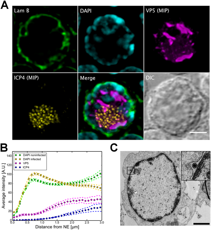
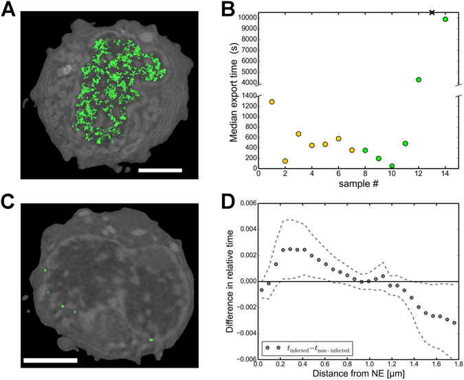
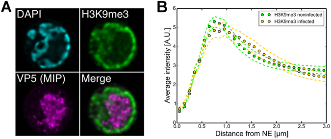
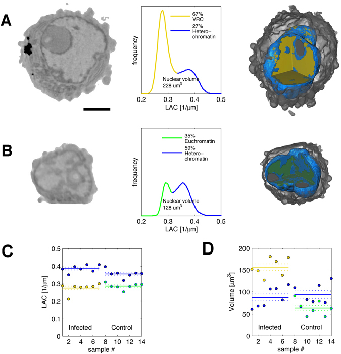
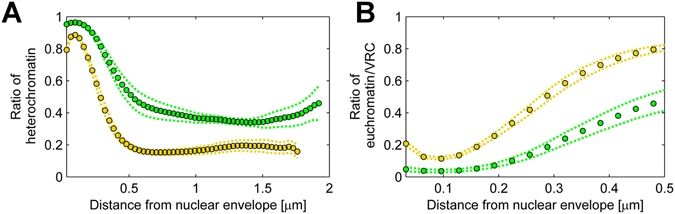
Various types of DNA viruses are known to elicit the formation of a large nuclear viral replication compartment and marginalization of the cell chromatin. We used three-dimensional soft x-ray tomography, confocal and electron microscopy, combined with numerical modelling of capsid diffusion to analyse the molecular organization of chromatin in herpes simplex virus 1 infection and its effect on the transport of progeny viral capsids to the nuclear envelope. Our data showed that the formation of the viral replication compartment at late infection resulted in the enrichment of heterochromatin in the nuclear periphery accompanied by the compaction of chromatin. Random walk modelling of herpes simplex virus 1-sized particles in a three-dimensional soft x-ray tomography reconstruction of an infected cell nucleus demonstrated that the peripheral, compacted chromatin restricts viral capsid diffusion, but due to interchromatin channels capsids are able to reach the nuclear envelope, the site of their nuclear egress.
已知各种类型的 DNA 病毒会引发大型核病毒复制 compartment 的形成,并使细胞染色质边缘化。我们使用三维软 X 射线断层扫描、共聚焦和电子显微镜,结合衣壳扩散的数值建模,分析单纯疱疹病毒 1 感染中染色质的分子组织及其对子代病毒衣壳向核膜运输的影响。我们的数据表明,晚期感染中病毒复制 compartment 的形成导致异染色质在核周富集,同时染色质浓缩。在感染细胞核的三维软 X 射线断层扫描重建中对单纯疱疹病毒 1 大小的粒子进行随机游走建模表明,外周致密的染色质限制了病毒衣壳的扩散,但由于染色质间通道的存在,衣壳能够到达核膜,即它们的核出口位点。