• 文献检索
  • 文档翻译
  • 深度研究
  • 学术资讯
  • Suppr Zotero 插件Zotero 插件
  • 邀请有礼
  • 套餐&价格
  • 历史记录
应用&插件
Suppr Zotero 插件Zotero 插件浏览器插件Mac 客户端Windows 客户端微信小程序
定价
高级版会员购买积分包购买API积分包
服务
文献检索文档翻译深度研究API 文档MCP 服务
关于我们
关于 Suppr公司介绍联系我们用户协议隐私条款
关注我们

Suppr 超能文献

核心技术专利:CN118964589B侵权必究
粤ICP备2023148730 号-1Suppr @ 2026

文献检索

告别复杂PubMed语法,用中文像聊天一样搜索,搜遍4000万医学文献。AI智能推荐,让科研检索更轻松。

立即免费搜索

文件翻译

保留排版,准确专业,支持PDF/Word/PPT等文件格式,支持 12+语言互译。

免费翻译文档

深度研究

AI帮你快速写综述,25分钟生成高质量综述,智能提取关键信息,辅助科研写作。

立即免费体验

基于三维T1rho成像的脑肿瘤相关水肿的鉴别

Differentiation of brain tumor-related edema based on 3D T1rho imaging.

作者信息

Villanueva-Meyer J E, Barajas R F, Mabray M C, Chen W, Shankaranarayanan A, Koon P, Barani I J, Tihan T, Cha S

机构信息

Department of Radiology and Biomedical Imaging, University of California San Francisco, San Francisco, CA, USA.

Department of Diagnostic Radiology, Oregon Health and Science University, Portland, OR, USA; Advanced Imaging Research Center, Oregon Health and Science University, Portland, OR, USA.

出版信息

Eur J Radiol. 2017 Jun;91:88-92. doi: 10.1016/j.ejrad.2017.03.022. Epub 2017 Mar 30.

DOI:10.1016/j.ejrad.2017.03.022
PMID:28629576
原文链接:https://pmc.ncbi.nlm.nih.gov/articles/PMC5563444/
Abstract

BACKGROUND AND PURPOSE

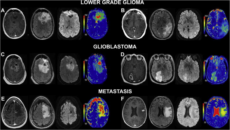
Cerebral edema associated with brain tumors is an important source of morbidity. Its type depends largely on the capillary ultra-structures of the histopathologic subtype of underlying brain tumor. The purpose of our study was to differentiate vasogenic edema associated with brain metastases and infiltrative edema related to diffuse gliomas using quantitative 3D T1 rho (T1ρ) imaging.

MATERIALS AND METHODS

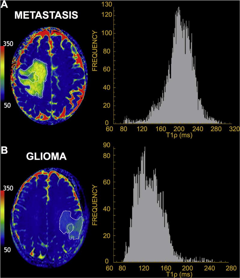
Preoperative MR examination including whole brain 3D T1ρ imaging was performed in 23 patients with newly diagnosed brain tumors (9 with metastasis, 8 with lower grade glioma, LGG, 6 with glioblastoma, GBM). Mean T1ρ values were measured in regions of peritumoral non-enhancing T2 signal hyperintensity, excluding both enhancing and necrotic or cystic component, and normal-appearing white matter.

RESULTS

Mean T1ρ values were significantly elevated in the vasogenic edema surrounding intracranial metastases when compared to the infiltrative edema associated with either LGG or GBM (p=0.02 and <0.01, respectively). No significant difference was noted between T1ρ values of infiltrative edema between LGG and GBM (p=0.84 and 0.96, respectively).

CONCLUSION

Our study demonstrates the feasibility and potential diagnostic role of T1ρ in the quantitative differentiation between edema related to intracranial metastases and gliomas and as a potentially complementary tool to standard MR techniques in further characterizing pathophysiology of vasogenic and infiltrative edema.

摘要

背景与目的

与脑肿瘤相关的脑水肿是发病的一个重要来源。其类型很大程度上取决于潜在脑肿瘤组织病理学亚型的毛细血管超微结构。我们研究的目的是使用定量三维T1ρ(T1ρ)成像来区分与脑转移瘤相关的血管源性水肿和与弥漫性胶质瘤相关的浸润性水肿。

材料与方法

对23例新诊断的脑肿瘤患者(9例转移瘤、8例低级别胶质瘤、6例胶质母细胞瘤)进行术前磁共振检查,包括全脑三维T1ρ成像。在瘤周非强化T2信号高强化区域测量平均T1ρ值,不包括强化、坏死或囊性成分以及外观正常的白质。

结果

与低级别胶质瘤或胶质母细胞瘤相关的浸润性水肿相比,颅内转移瘤周围血管源性水肿的平均T1ρ值显著升高(分别为p = 0.02和<0.01)。低级别胶质瘤和胶质母细胞瘤的浸润性水肿T1ρ值之间无显著差异(分别为p = 0.84和0.96)。

结论

我们的研究证明了T1ρ在定量区分颅内转移瘤和胶质瘤相关水肿方面的可行性和潜在诊断作用,并且作为一种潜在的补充工具,可用于进一步表征血管源性和浸润性水肿的病理生理学,辅助标准磁共振技术。

https://cdn.ncbi.nlm.nih.gov/pmc/blobs/5837/5563444/257f4290cf3d/nihms893704f3.jpg
https://cdn.ncbi.nlm.nih.gov/pmc/blobs/5837/5563444/1b2d12101259/nihms893704f1.jpg
https://cdn.ncbi.nlm.nih.gov/pmc/blobs/5837/5563444/dd927f72728e/nihms893704f2.jpg
https://cdn.ncbi.nlm.nih.gov/pmc/blobs/5837/5563444/257f4290cf3d/nihms893704f3.jpg
https://cdn.ncbi.nlm.nih.gov/pmc/blobs/5837/5563444/1b2d12101259/nihms893704f1.jpg
https://cdn.ncbi.nlm.nih.gov/pmc/blobs/5837/5563444/dd927f72728e/nihms893704f2.jpg
https://cdn.ncbi.nlm.nih.gov/pmc/blobs/5837/5563444/257f4290cf3d/nihms893704f3.jpg

相似文献

1
Differentiation of brain tumor-related edema based on 3D T1rho imaging.基于三维T1rho成像的脑肿瘤相关水肿的鉴别
Eur J Radiol. 2017 Jun;91:88-92. doi: 10.1016/j.ejrad.2017.03.022. Epub 2017 Mar 30.
2
Quantitative apparent diffusion coefficients in the characterization of brain tumors and associated peritumoral edema.定量表观扩散系数在脑肿瘤及相关瘤周水肿特征描述中的应用
Acta Radiol. 2009 Jul;50(6):682-9. doi: 10.1080/02841850902933123.
3
Differentiation between vasogenic-edema versus tumor-infiltrative area in patients with glioblastoma during bevacizumab therapy: a longitudinal MRI study.贝伐单抗治疗期间胶质母细胞瘤患者血管源性水肿与肿瘤浸润区域的鉴别:一项纵向MRI研究
Eur J Radiol. 2014 Jul;83(7):1250-1256. doi: 10.1016/j.ejrad.2014.03.026. Epub 2014 Apr 12.
4
Quantitative apparent diffusion coefficients and T2 relaxation times in characterizing contrast enhancing brain tumors and regions of peritumoral edema.定量表观扩散系数和T2弛豫时间在鉴别强化脑肿瘤及瘤周水肿区域中的应用
J Magn Reson Imaging. 2005 Jun;21(6):701-8. doi: 10.1002/jmri.20335.
5
Differentiation of pure vasogenic edema and tumor-infiltrated edema in patients with peritumoral edema by analyzing the relationship of axial and radial diffusivities on 3.0T MRI.通过分析3.0T磁共振成像(MRI)上轴向和径向扩散率之间的关系,鉴别肿瘤周围水肿患者的单纯血管源性水肿和肿瘤浸润性水肿。
Clin Neurol Neurosurg. 2013 Aug;115(8):1366-70. doi: 10.1016/j.clineuro.2012.12.031. Epub 2013 Jan 23.
6
The differences of water diffusion between brain tissue infiltrated by tumor and peritumoral vasogenic edema.肿瘤浸润脑组织与瘤周血管源性水肿之间水扩散的差异。
Clin Imaging. 2009 Mar-Apr;33(2):96-101. doi: 10.1016/j.clinimag.2008.06.035.
7
Measurements of diagnostic examination performance using quantitative apparent diffusion coefficient and proton MR spectroscopic imaging in the preoperative evaluation of tumor grade in cerebral gliomas.使用定量表观扩散系数和质子磁共振波谱成像对脑胶质瘤术前肿瘤分级进行诊断检查性能的测量。
Eur J Radiol. 2011 Nov;80(2):462-70. doi: 10.1016/j.ejrad.2010.07.017. Epub 2010 Aug 13.
8
Advanced diffusion MRI provides evidence for altered axonal microstructure and gradual peritumoral infiltration in GBM in comparison to brain metastases.高级弥散 MRI 为胶质母细胞瘤与脑转移瘤相比,存在轴突微观结构改变和逐渐发生的肿瘤周围浸润提供了证据。
Clin Neuroradiol. 2024 Sep;34(3):703-711. doi: 10.1007/s00062-024-01416-0. Epub 2024 Apr 29.
9
On differentiation between vasogenic edema and non-enhancing tumor in high-grade glioma patients using a support vector machine classifier based upon pre and post-surgery MRI images.基于术前和术后 MRI 图像的支持向量机分类器在高级别胶质瘤患者中对血管源性水肿和无强化肿瘤的鉴别。
Eur J Radiol. 2018 Sep;106:199-208. doi: 10.1016/j.ejrad.2018.07.018. Epub 2018 Jul 20.
10
Peritumoral brain regions in gliomas and meningiomas: investigation with isotropic diffusion-weighted MR imaging and diffusion-tensor MR imaging.胶质瘤和脑膜瘤周围脑区:采用各向同性扩散加权磁共振成像和扩散张量磁共振成像进行研究
Radiology. 2004 Aug;232(2):451-60. doi: 10.1148/radiol.2322030959. Epub 2004 Jun 23.

引用本文的文献

1
Adaptive Radiation Therapy for Head and Neck Cancer.头颈部癌的自适应放射治疗
ArXiv. 2025 Aug 1:arXiv:2508.00651v1.
2
Machine learning-based nomogram for distinguishing between supratentorial extraventricular ependymoma and supratentorial glioblastoma.基于机器学习的列线图用于鉴别幕上脑室外室管膜瘤和幕上胶质母细胞瘤
Front Oncol. 2024 Sep 9;14:1443913. doi: 10.3389/fonc.2024.1443913. eCollection 2024.
3
Amide proton transfer-weighted and arterial spin labeling imaging may improve differentiation between high-grade glioma recurrence and radiation-induced brain injury.

本文引用的文献

1
In vivo quantitative whole-brain T1 rho MRI of multiple sclerosis.多发性硬化症的体内全脑定量T1ρ磁共振成像
J Magn Reson Imaging. 2015 Dec;42(6):1623-30. doi: 10.1002/jmri.24954. Epub 2015 May 30.
2
T1rho MRI and CSF biomarkers in diagnosis of Alzheimer's disease.T1rho磁共振成像与脑脊液生物标志物在阿尔茨海默病诊断中的应用
Neuroimage Clin. 2015 Feb 26;7:598-604. doi: 10.1016/j.nicl.2015.02.016. eCollection 2015.
3
Brain abnormalities in bipolar disorder detected by quantitative T1ρ mapping.通过定量T1ρ成像检测双相情感障碍中的脑异常。
酰胺质子转移加权成像和动脉自旋标记成像可能会改善高级别胶质瘤复发与放射性脑损伤之间的鉴别诊断。
Heliyon. 2024 Jun 7;10(11):e32699. doi: 10.1016/j.heliyon.2024.e32699. eCollection 2024 Jun 15.
4
Magnetic resonance relaxometry in quantitative imaging of brain gliomas: A literature review.磁共振弛豫定量成像在脑胶质瘤中的应用:文献综述。
Neuroradiol J. 2024 Jun;37(3):267-275. doi: 10.1177/19714009231173100. Epub 2023 May 3.
5
Targeted Arginine Metabolism Therapy: A Dilemma in Glioma Treatment.靶向精氨酸代谢疗法:胶质瘤治疗中的一个困境
Front Oncol. 2022 Jul 11;12:938847. doi: 10.3389/fonc.2022.938847. eCollection 2022.
6
Quantitative T1ρ MRI of the Head and Neck Discriminates Carcinoma and Benign Hyperplasia in the Nasopharynx.头颈部定量 T1ρ MRI 鉴别鼻咽癌中的癌与良性增生。
AJNR Am J Neuroradiol. 2020 Dec;41(12):2339-2344. doi: 10.3174/ajnr.A6828. Epub 2020 Oct 29.
7
T magnetic resonance fingerprinting.T 磁共振指纹成像。
NMR Biomed. 2020 May;33(5):e4284. doi: 10.1002/nbm.4284. Epub 2020 Mar 3.
8
Histogram analysis of absolute cerebral blood volume map can distinguish glioblastoma from solitary brain metastasis.绝对脑血容量图的直方图分析可区分胶质母细胞瘤与孤立性脑转移瘤。
Medicine (Baltimore). 2019 Oct;98(42):e17515. doi: 10.1097/MD.0000000000017515.
9
Brain T1ρ mapping for grading and IDH1 gene mutation detection of gliomas: a preliminary study.脑 T1ρ 图谱在胶质瘤分级和 IDH1 基因突变检测中的应用:一项初步研究。
J Neurooncol. 2019 Jan;141(1):245-252. doi: 10.1007/s11060-018-03033-7. Epub 2018 Nov 9.
Mol Psychiatry. 2015 Feb;20(2):201-6. doi: 10.1038/mp.2014.157. Epub 2015 Jan 6.
4
Magnetization transfer and adiabatic T1ρ MRI reveal abnormalities in normal-appearing white matter of subjects with multiple sclerosis.磁化传递和绝热T1ρ磁共振成像揭示了多发性硬化症患者正常外观白质中的异常。
Mult Scler. 2014 Jul;20(8):1066-73. doi: 10.1177/1352458513515084. Epub 2013 Dec 12.
5
In vivo whole-brain T1-rho mapping across adulthood: normative values and age dependence.成年期全脑T1-ρ图谱的活体研究:正常参考值及年龄依赖性
J Magn Reson Imaging. 2014 Aug;40(2):376-82. doi: 10.1002/jmri.24358. Epub 2013 Nov 13.
6
Toward determining the lifetime occurrence of metastatic brain tumors estimated from 2007 United States cancer incidence data.从 2007 年美国癌症发病率数据估算转移性脑肿瘤的终生发病情况。
Neuro Oncol. 2012 Sep;14(9):1171-7. doi: 10.1093/neuonc/nos152. Epub 2012 Aug 16.
7
Detecting activity-evoked pH changes in human brain.检测人脑活动诱发的 pH 值变化。
Proc Natl Acad Sci U S A. 2012 May 22;109(21):8270-3. doi: 10.1073/pnas.1205902109. Epub 2012 May 7.
8
Imaging the extracellular pH of tumors by MRI after injection of a single cocktail of T1 and T2 contrast agents.注射单一鸡尾酒 T1 和 T2 对比剂后通过 MRI 对肿瘤细胞外 pH 值进行成像。
NMR Biomed. 2011 Dec;24(10):1380-91. doi: 10.1002/nbm.1701. Epub 2011 May 23.
9
T1rho (T1ρ) MR imaging in Alzheimer's disease and Parkinson's disease with and without dementia.阿尔茨海默病和帕金森病伴或不伴痴呆的 T1rho(T1ρ)磁共振成像。
J Neurol. 2011 Mar;258(3):380-5. doi: 10.1007/s00415-010-5762-6. Epub 2010 Oct 7.
10
Estimation of the onset time of cerebral ischemia using T1rho and T2 MRI in rats.利用 T1rho 和 T2MRI 估计大鼠脑缺血的发作时间。
Stroke. 2010 Oct;41(10):2335-40. doi: 10.1161/STROKEAHA.110.587394. Epub 2010 Sep 2.