• 文献检索
  • 文档翻译
  • 深度研究
  • 学术资讯
  • Suppr Zotero 插件Zotero 插件
  • 邀请有礼
  • 套餐&价格
  • 历史记录
应用&插件
Suppr Zotero 插件Zotero 插件浏览器插件Mac 客户端Windows 客户端微信小程序
定价
高级版会员购买积分包购买API积分包
服务
文献检索文档翻译深度研究API 文档MCP 服务
关于我们
关于 Suppr公司介绍联系我们用户协议隐私条款
关注我们

Suppr 超能文献

核心技术专利:CN118964589B侵权必究
粤ICP备2023148730 号-1Suppr @ 2026

文献检索

告别复杂PubMed语法,用中文像聊天一样搜索,搜遍4000万医学文献。AI智能推荐,让科研检索更轻松。

立即免费搜索

文件翻译

保留排版,准确专业,支持PDF/Word/PPT等文件格式,支持 12+语言互译。

免费翻译文档

深度研究

AI帮你快速写综述,25分钟生成高质量综述,智能提取关键信息,辅助科研写作。

立即免费体验

无创冠状动脉波强度分析。

Non-invasive coronary wave intensity analysis.

作者信息

Broyd Christopher J, Rigo Fausto, Davies Justin

机构信息

Imperial College London, London, UK.

National Heart and Lung Institute, Hammersmith Hospital, Du Cane Road, London, W12 0HS, UK.

出版信息

Int J Cardiovasc Imaging. 2017 Jul;33(7):1061-1068. doi: 10.1007/s10554-017-1185-0. Epub 2017 Jun 19.

DOI:10.1007/s10554-017-1185-0
PMID:28631107
原文链接:https://pmc.ncbi.nlm.nih.gov/articles/PMC5489579/
Abstract

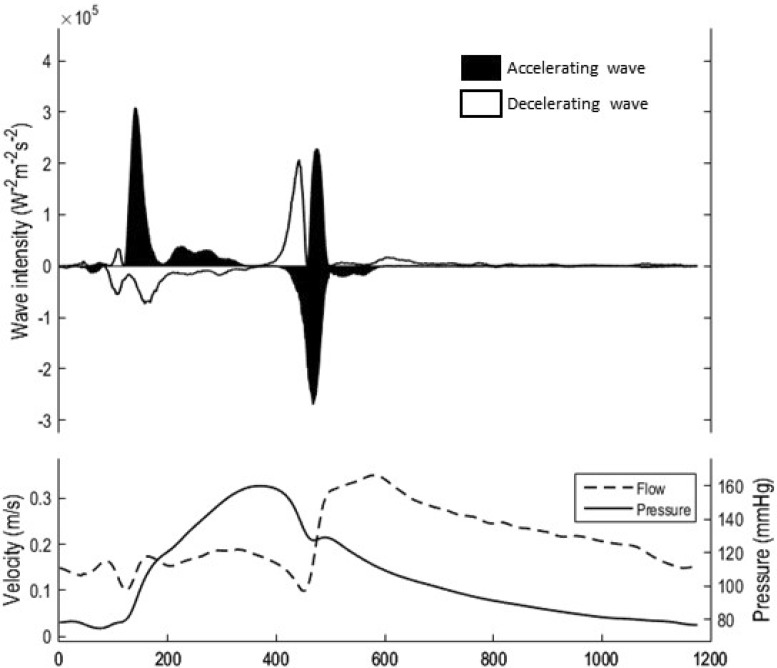
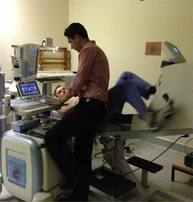
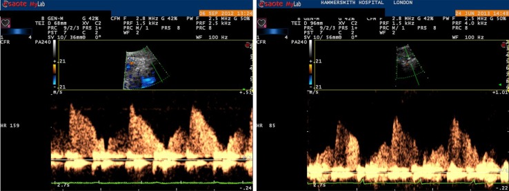
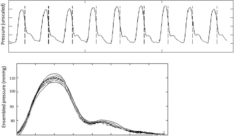
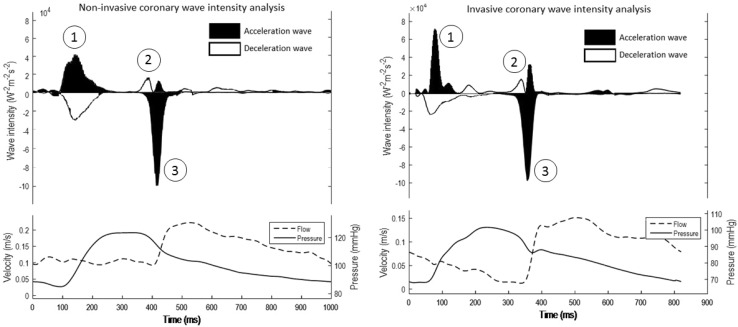
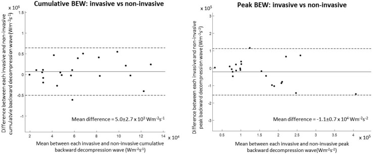
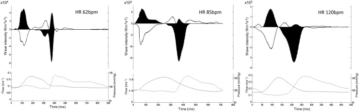
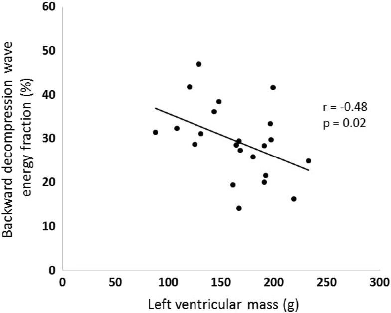
Wave intensity analysis is calculated from simultaneously acquired measures of pressure and flow. Its mathematical computation produces a profile that provides quantitative information on the energy exchange driving blood flow acceleration and deceleration. Within the coronary circulation it has proven most useful in describing the wave that originates from the myocardium and that is responsible for driving the majority of coronary flow, labelled the backward decompression wave. Whilst this wave has demonstrated valuable insights into the pathogenic processes of a number of disease states, its measurement is hampered by its invasive necessity. However, recent work has used transthoracic echocardiography and an established measures of central aortic pressure to produce coronary flow velocity and pressure waveforms respectively. This has allowed a non-invasive measure of coronary wave intensity analysis, and in particular the backward decompression wave, to be calculated. It is anticipated that this will allow this tool to become more applicable and widespread, ultimately moving it from the research to the clinical domain.

摘要

波强度分析是根据同时采集的压力和流量测量值计算得出的。其数学计算产生一个剖面图,该剖面图提供了关于驱动血流加速和减速的能量交换的定量信息。在冠状动脉循环中,它已被证明在描述源自心肌并负责驱动大部分冠状动脉血流的波(标记为反向减压波)方面最为有用。虽然这个波已经为许多疾病状态的致病过程提供了有价值的见解,但其测量因其侵入性的必要性而受到阻碍。然而,最近的研究使用经胸超声心动图和一种既定的中心主动脉压测量方法,分别产生冠状动脉流速和压力波形。这使得能够对冠状动脉波强度分析,特别是反向减压波进行非侵入性测量。预计这将使该工具更具适用性和广泛应用,最终将其从研究领域转移到临床领域。

https://cdn.ncbi.nlm.nih.gov/pmc/blobs/3fab/5489579/ce4173604576/10554_2017_1185_Fig8_HTML.jpg
https://cdn.ncbi.nlm.nih.gov/pmc/blobs/3fab/5489579/2547011617a3/10554_2017_1185_Fig1_HTML.jpg
https://cdn.ncbi.nlm.nih.gov/pmc/blobs/3fab/5489579/f3e5d8e01a9c/10554_2017_1185_Fig2_HTML.jpg
https://cdn.ncbi.nlm.nih.gov/pmc/blobs/3fab/5489579/a79e7bdb1cd8/10554_2017_1185_Fig3_HTML.jpg
https://cdn.ncbi.nlm.nih.gov/pmc/blobs/3fab/5489579/b379086fd4b8/10554_2017_1185_Fig4_HTML.jpg
https://cdn.ncbi.nlm.nih.gov/pmc/blobs/3fab/5489579/8b6afc2ca719/10554_2017_1185_Fig5_HTML.jpg
https://cdn.ncbi.nlm.nih.gov/pmc/blobs/3fab/5489579/93594d414a4d/10554_2017_1185_Fig6_HTML.jpg
https://cdn.ncbi.nlm.nih.gov/pmc/blobs/3fab/5489579/6798ff8b982c/10554_2017_1185_Fig7_HTML.jpg
https://cdn.ncbi.nlm.nih.gov/pmc/blobs/3fab/5489579/ce4173604576/10554_2017_1185_Fig8_HTML.jpg

相似文献

1
Non-invasive coronary wave intensity analysis.无创冠状动脉波强度分析。
Int J Cardiovasc Imaging. 2017 Jul;33(7):1061-1068. doi: 10.1007/s10554-017-1185-0. Epub 2017 Jun 19.
2
Assessment of left anterior descending artery stenosis of intermediate severity by fractional flow reserve, instantaneous wave-free ratio, and non-invasive coronary flow reserve.通过血流储备分数、瞬时无波比值和无创冠状动脉血流储备评估中度左前降支动脉狭窄
Int J Cardiovasc Imaging. 2017 Jul;33(7):999-1007. doi: 10.1007/s10554-016-1000-3. Epub 2016 Oct 17.
3
Cardiac PET imaging for the detection and monitoring of coronary artery disease and microvascular health.心脏 PET 成像在冠状动脉疾病和微血管健康的检测和监测中的应用。
JACC Cardiovasc Imaging. 2010 Jun;3(6):623-40. doi: 10.1016/j.jcmg.2010.04.007.
4
Invasive coronary physiology for assessing intermediate lesions.用于评估中等病变的有创冠状动脉生理学
Circ Cardiovasc Interv. 2015 Feb;8(2):e001942. doi: 10.1161/CIRCINTERVENTIONS.114.001942.
5
Targeting the dominant mechanism of coronary microvascular dysfunction with intracoronary physiology tests.通过冠状动脉内生理学测试针对冠状动脉微血管功能障碍的主要机制。
Int J Cardiovasc Imaging. 2017 Jul;33(7):1041-1059. doi: 10.1007/s10554-017-1136-9. Epub 2017 May 13.
6
Exploring Coronary Circulatory Response to Stenosis and Its Association With Invasive Physiologic Indexes Using Absolute Myocardial Blood Flow and Coronary Pressure.探讨使用绝对心肌血流和冠状动脉压力评估狭窄病变时冠状动脉循环反应及其与有创生理学指标的相关性。
Circulation. 2017 Nov 7;136(19):1798-1808. doi: 10.1161/CIRCULATIONAHA.117.029911. Epub 2017 Aug 29.
7
Revisiting the Optimal Fractional Flow Reserve and Instantaneous Wave-Free Ratio Thresholds for Predicting the Physiological Significance of Coronary Artery Disease.重新探讨用于预测冠状动脉疾病生理学意义的最佳血流储备分数和瞬时无波比值阈值。
Circ Cardiovasc Interv. 2018 Dec;11(12):e007041. doi: 10.1161/CIRCINTERVENTIONS.118.007041.
8
Resting Coronary Flow Varies With Normal Cardiac Catheter Laboratory Stimuli.静息冠状动脉血流随心脏导管实验室的正常刺激而变化。
Cardiovasc Revasc Med. 2019 Aug;20(8):669-673. doi: 10.1016/j.carrev.2018.10.010. Epub 2018 Oct 15.
9
The reasons why fractional flow reserve and instantaneous wave-free ratio are similar using wave separation analysis.采用波分离分析时,分数流量储备和瞬时无波比相似的原因。
BMC Cardiovasc Disord. 2021 Jan 25;21(1):48. doi: 10.1186/s12872-021-01855-4.
10
Peripheral Reactive Hyperemia Index and Coronary Microvascular Function in Women With no Obstructive CAD: The iPOWER Study.无阻塞性 CAD 女性的外周反应性充血指数与冠状动脉微血管功能:iPOWER 研究。
JACC Cardiovasc Imaging. 2016 Apr;9(4):411-7. doi: 10.1016/j.jcmg.2016.02.005.

引用本文的文献

1
The Use of Maximum Entropy to Enhance Wave Intensity Analysis: An Application to Coronary Arteries in Hypertrophic Obstructive Cardiomyopathy.利用最大熵增强波强度分析:在肥厚性梗阻性心肌病冠状动脉中的应用
Front Cardiovasc Med. 2021 Aug 27;8:701267. doi: 10.3389/fcvm.2021.701267. eCollection 2021.
2
Cardiovascular imaging 2017 in the International Journal of Cardiovascular Imaging.《国际心血管影像杂志》2017年心血管成像相关内容
Int J Cardiovasc Imaging. 2018 Jun;34(6):833-848. doi: 10.1007/s10554-018-1349-6.
3
Whence we came, whither we go?

本文引用的文献

1
Wave intensity analysis and its application to the coronary circulation.波强度分析及其在冠状动脉循环中的应用。
Glob Cardiol Sci Pract. 2017 Mar 31;2017(1):e201705. doi: 10.21542/gcsp.2017.5.
2
Estimation of coronary wave intensity analysis using noninvasive techniques and its application to exercise physiology.利用无创技术估计冠状动脉波强度分析及其在运动生理学中的应用。
Am J Physiol Heart Circ Physiol. 2016 Mar 1;310(5):H619-27. doi: 10.1152/ajpheart.00575.2015. Epub 2015 Dec 18.
3
Myocardial ischemia in aortic stenosis: insights from arterial pulse-wave dynamics after percutaneous aortic valve replacement.
我们从何处来,又将向何处去?
Int J Cardiovasc Imaging. 2017 Jul;33(7):957-959. doi: 10.1007/s10554-017-1166-3.
主动脉瓣狭窄患者心肌缺血:经皮主动脉瓣置换术后动脉脉搏波动力学的新认识。
Trends Cardiovasc Med. 2013 Aug;23(6):185-91. doi: 10.1016/j.tcm.2012.12.001. Epub 2013 Feb 8.
4
Coronary flow reserve during dipyridamole stress echocardiography predicts mortality.在双嘧达莫负荷超声心动图期间的冠状动脉血流储备预测死亡率。
JACC Cardiovasc Imaging. 2012 Nov;5(11):1079-85. doi: 10.1016/j.jcmg.2012.08.007.
5
Synergistic adaptations to exercise in the systemic and coronary circulations that underlie the warm-up angina phenomenon.运动对全身和冠状动脉循环的协同适应,是热身心绞痛现象的基础。
Circulation. 2012 Nov 27;126(22):2565-74. doi: 10.1161/CIRCULATIONAHA.112.094292. Epub 2012 Nov 1.
6
Improvement in coronary blood flow velocity with acute biventricular pacing is predominantly due to an increase in a diastolic backward-travelling decompression (suction) wave.急性双心室起搏时冠状动脉血流速度的改善主要归因于舒张期逆行减压(抽吸)波的增加。
Circulation. 2012 Sep 11;126(11):1334-44. doi: 10.1161/CIRCULATIONAHA.111.075606. Epub 2012 Aug 1.
7
Evaluation of a novel sphygmomanometer, which estimates central aortic blood pressure from analysis of brachial artery suprasystolic pressure waves.评估一种新型的血压计,该血压计通过分析肱动脉超射压力波来估算中心主动脉血压。
J Hypertens. 2012 Sep;30(9):1743-50. doi: 10.1097/HJH.0b013e3283567b94.
8
Arterial pulse wave dynamics after percutaneous aortic valve replacement: fall in coronary diastolic suction with increasing heart rate as a basis for angina symptoms in aortic stenosis.经皮主动脉瓣置换术后动脉脉搏波动力学:心率增加时冠状动脉舒张压抽吸下降,是主动脉瓣狭窄心绞痛症状的基础。
Circulation. 2011 Oct 4;124(14):1565-72. doi: 10.1161/CIRCULATIONAHA.110.011916. Epub 2011 Sep 12.
9
Validation of a brachial cuff-based method for estimating central systolic blood pressure.验证一种基于肱动脉袖带的方法来估计中心收缩压。
Hypertension. 2011 Nov;58(5):825-32. doi: 10.1161/HYPERTENSIONAHA.111.176313. Epub 2011 Sep 12.
10
A new oscillometric method for pulse wave analysis: comparison with a common tonometric method.一种新的脉搏波分析示波法:与常用的测振法比较。
J Hum Hypertens. 2010 Aug;24(8):498-504. doi: 10.1038/jhh.2010.27. Epub 2010 Mar 18.