Rösch Frank, Herzog Hans, Qaim Syed M
Institute of Nuclear Chemistry, Johannes Gutenberg University Mainz, Mainz D-55126, Germany.
Institute of Neuroscience and Medicine (INM), INM-4 (Physics of Medical Imaging), Research Center Jülich, Jülich D-52425, Germany.
Pharmaceuticals (Basel). 2017 Jun 20;10(2):56. doi: 10.3390/ph10020056.
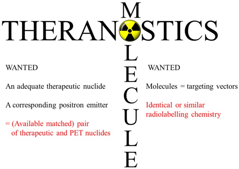
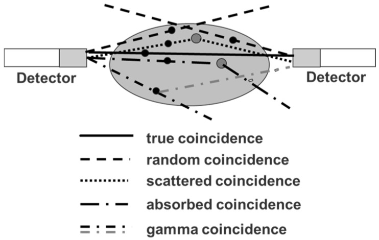
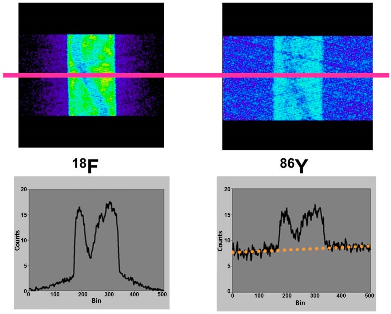
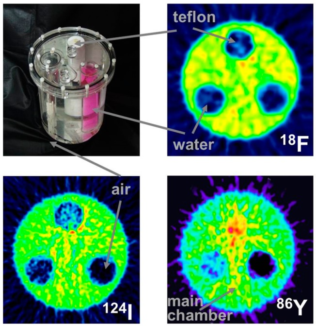
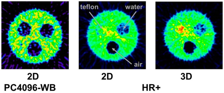
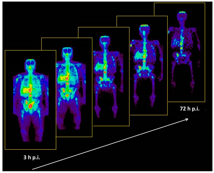
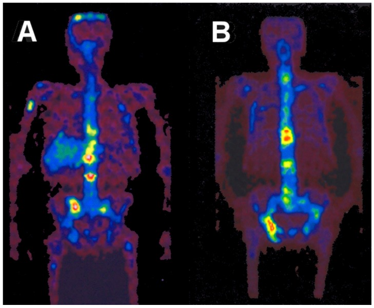
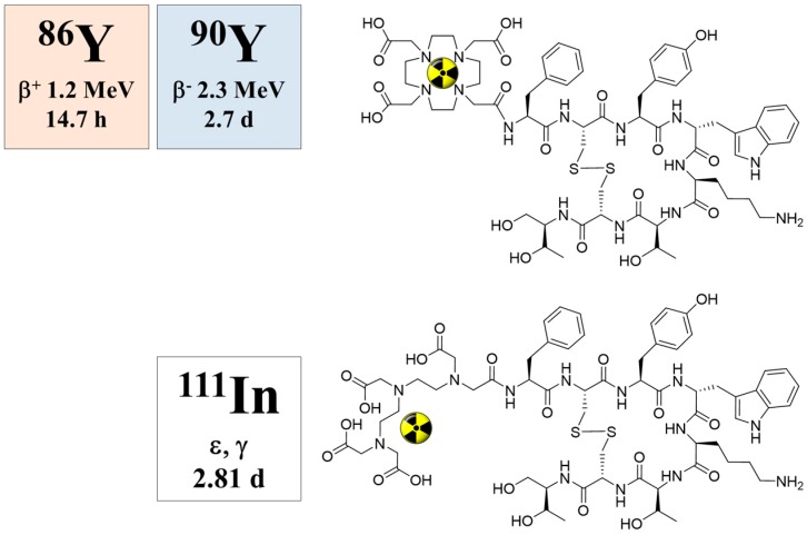
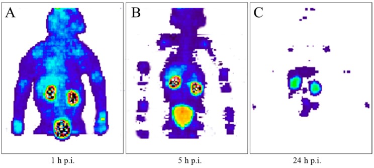
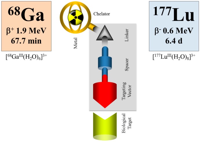
In the context of radiopharmacy and molecular imaging, the concept of theranostics entails a therapy-accompanying diagnosis with the aim of a patient-specific treatment. Using the adequate diagnostic radiopharmaceutical, the disease and the state of the disease are verified for an individual patient. The other way around, it verifies that the radiopharmaceutical in hand represents a target-specific and selective molecule: the "best one" for that individual patient. Transforming diagnostic imaging into quantitative dosimetric information, the optimum radioactivity (expressed in maximum radiation dose to the target tissue and tolerable dose to healthy organs) of the adequate radiotherapeutical is applied to that individual patient. This theranostic approach in nuclear medicine is traced back to the first use of the radionuclide pair Y/Y, which allowed a combination of PET and internal radiotherapy. Whereas the β-emitting therapeutic radionuclide Y (t½ = 2.7 d) had been available for a long time via the Sr/Y generator system, the β⁺ emitter Y (t½ = 14.7 h) had to be developed for medical application. A brief outline of the various aspects of radiochemical and nuclear development work (nuclear data, cyclotron irradiation, chemical processing, quality control, etc.) is given. In parallel, the paper discusses the methodology introduced to quantify molecular imaging of Y-labelled compounds in terms of multiple and long-term PET recordings. It highlights the ultimate goal of radiotheranostics, namely to extract the radiation dose of the analogue Y-labelled compound in terms of mGy or mSv per MBq Y injected. Finally, the current and possible future development of theranostic approaches based on different PET and therapy nuclides is discussed.
在放射性药物学和分子成像领域,治疗诊断学的概念是指伴随治疗的诊断,旨在实现针对患者个体的治疗。使用适当的诊断性放射性药物,可针对个体患者验证疾病及其病情状态。反之,也可验证手头的放射性药物是否是一种靶向特异性且具有选择性的分子:即对该个体患者而言的“最佳药物”。将诊断成像转化为定量剂量信息后,将适当的放射治疗药物的最佳放射性活度(以对靶组织的最大辐射剂量和对健康器官的可耐受剂量表示)应用于该个体患者。核医学中的这种治疗诊断方法可追溯到放射性核素对Y/Y的首次使用,它实现了正电子发射断层扫描(PET)与内放射治疗的结合。虽然发射β射线的治疗性放射性核素Y(半衰期t½ = 2.7天)可通过Sr/Y发生器系统长期获得,但发射β⁺射线的Y(半衰期t½ = 14.7小时)必须研发用于医学应用。本文简要概述了放射化学和核技术开发工作的各个方面(核数据、回旋加速器辐照、化学处理、质量控制等)。同时,本文还讨论了为通过多次长期PET记录来量化Y标记化合物的分子成像而引入的方法。它强调了放射治疗诊断学的最终目标,即以每注射1MBq Y的毫戈瑞(mGy)或毫希沃特(mSv)为单位,推算出类似Y标记化合物的辐射剂量。最后,本文讨论了基于不同PET和治疗核素的治疗诊断方法的当前及可能的未来发展。