Wei Xiaolong, Sun Yudong, Wu Yani, Zhu Jiang, Gao Bin, Yan Han, Zhao Zhiqing, Zhou Jian, Jing Zaiping
Department of Vascular Surgery, Changhai Hospital, Second Military Medical University, 168 Changhai Road, Shanghai, 200433, China.
Company 8, Cadet brigade, Second Military Medical University, Shanghai, China.
BMC Cardiovasc Disord. 2017 Jun 20;17(1):162. doi: 10.1186/s12872-017-0588-0.
This study aimed to assessed whether Talin-1 is involved in the pathogenesis of aortic dissection via regulating vascular smooth muscle cell (VSMC) biological function.
Human aortic samples were obtained from organ donors who died from nonvascular diseases as normal controls and from patients undergoing surgical repair of thoracic aortic dissection. The expression level and distribution of Talin-1 were detected using westernblot analysis and immunohistochemistry in each sample. We inhibited the expression of Talin-1 via RNA interference in VSMCs. VSMC proliferation was detected by Cell-counting Kit-8 analyses. Scratch test and flow cytometry were used to identify the migration and apoptosis ability. Antibody microarray analysis and qRT-PCR were used to detect some protein and mRNA changes which were induced by Talin-1 downregulation.
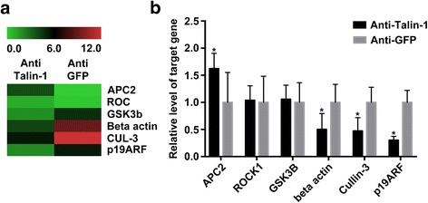
Talin-1 was significantly downregulated in the media of aortic dissection samples compared with controls (P < 0.05). Talin-1 knockdown significantly induced VSMC proliferation and migration in vitro. Proteins which involved in cell cycle can be regulated by downregulating Talin-1. Down regulation of Talin-1 can significanly increased the expression of anaphase-promoting complex subunit 2 (APC2) and decreased p19 alternative reading frame (p19ARF), Cullin-3, and beta actin's expression.
Talin-1 induces VSMCs proliferation and migration. It downregulated in aortic dissection, which might play a potential role in the development of aortic dissection.
本研究旨在评估踝蛋白-1(Talin-1)是否通过调节血管平滑肌细胞(VSMC)生物学功能参与主动脉夹层的发病机制。
从死于非血管疾病的器官捐献者获取人主动脉样本作为正常对照,以及从接受胸主动脉夹层手术修复的患者获取样本。采用蛋白质免疫印迹分析和免疫组织化学检测每个样本中Talin-1的表达水平和分布。我们通过RNA干扰在血管平滑肌细胞中抑制Talin-1的表达。采用细胞计数试剂盒-8分析检测血管平滑肌细胞增殖。划痕试验和流式细胞术用于鉴定迁移和凋亡能力。抗体微阵列分析和qRT-PCR用于检测Talin-1下调诱导的一些蛋白质和mRNA变化。
与对照组相比,主动脉夹层样本中膜层的Talin-1显著下调(P < 0.05)。敲低Talin-1在体外显著诱导血管平滑肌细胞增殖和迁移。下调Talin-1可调节参与细胞周期的蛋白质。Talin-1的下调可显著增加后期促进复合物亚基2(APC2)的表达,并降低p19可变阅读框(p19ARF)、Cullin-3和β-肌动蛋白的表达。
Talin-1诱导血管平滑肌细胞增殖和迁移。它在主动脉夹层中下调,这可能在主动脉夹层的发生发展中起潜在作用。