Lee Young-Wook, Lee Hyung, Chung In-Sung, Yi Hyon-Ah
Department of Neurology Brain Research Institute Department of Occupational and Environmental Medicine, Keimyung University School of Medicine, Daegu, South Korea.
Medicine (Baltimore). 2017 Jun;96(25):e7286. doi: 10.1097/MD.0000000000007286.
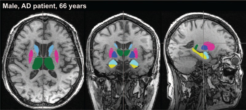
The relationship between postural instability and subcortical structure in AD has received less attention. The aims of this study were to assess whether there are differences in the ability to control balance between Alzheimer's disease (AD) and controls, and to investigate the association between subcortical gray matter volumes and postural instability in AD.We enrolled 107 consecutive AD patients and 37 controls. All participants underwent detailed neuropsychological evaluations, T1-weighted MRI at 3 T, and posture assessment using computerized dynamic posturography. We segmented the volumes of 6 subcortical structures of the amygdala, thalamus, caudate nucleus, putamen, globus pallidus and nucleus accumbens, and of hippocampus, using the FMRIBs integrated registration and segmentation tool.All subcortical structures, except for the globus pallidus, were smaller in AD compared with controls on adjusting for age and gender. Falling frequencies in unilateral stance test (UST) and composite scores in sensory organization test (SOT) were worse in AD than in controls. The motor control test did not reveal any differences between groups. On subgroup analyses in AD, the groups with poor performance in UST or SOT exhibited significantly reduced nucleus accumbens and putamen volumes, and nucleus accumbens volume, respectively. The smaller volume of the nucleus accumbens was associated with postural instability in AD (OR [95% CI] 17.847 [2.59-122.80] for UST, 42.827[6.06-302.47] for SOT, all P < .05).AD patients exhibited reduced ability to control balance compared with controls, and this postural instability was associated with nucleus accumbens volume loss. Furthermore, cognitive dysfunction was more prominent in the group with severe postural instability.
阿尔茨海默病(AD)中姿势不稳与皮质下结构之间的关系较少受到关注。本研究的目的是评估AD患者与对照组在平衡控制能力上是否存在差异,并研究AD患者皮质下灰质体积与姿势不稳之间的关联。我们连续纳入了107例AD患者和37例对照。所有参与者均接受了详细的神经心理学评估、3T的T1加权磁共振成像(MRI)以及使用计算机化动态姿势描记法进行的姿势评估。我们使用FMRIB综合配准和分割工具对杏仁核、丘脑、尾状核、壳核、苍白球、伏隔核以及海马的6个皮质下结构的体积进行了分割。在调整年龄和性别后,除苍白球外,AD患者所有皮质下结构的体积均小于对照组。AD患者在单腿站立试验(UST)中的跌倒频率和感觉组织试验(SOT)中的综合得分均比对照组差。运动控制测试未显示出两组之间存在任何差异。在AD患者的亚组分析中,UST或SOT表现较差的组分别显示伏隔核和壳核体积以及伏隔核体积显著减小。伏隔核体积较小与AD患者的姿势不稳相关(UST的比值比[95%可信区间]为17.847[2.59 - 122.80],SOT为42.827[6.06 - 302.47],均P<0.05)。与对照组相比,AD患者控制平衡的能力下降,且这种姿势不稳与伏隔核体积减小有关。此外,姿势不稳严重的组认知功能障碍更为突出。