Grunert Peter, Moriguchi Yu, Grossbard Brian P, Ricart Arbona Rodolfo J, Bonassar Lawrence J, Härtl Roger
Department of Neurological Surgery, Weill Cornell Medicine, New York-Presbyterian Hospital, Weill Cornell Brain and Spine Institute|, 525 East 68th Street, Box 99, New York, NY, 10065, USA.
Department of Neurological Surgery, Swedish Neuroscience Institute, Seattle, WA, USA.
BMC Vet Res. 2017 Jun 23;13(1):193. doi: 10.1186/s12917-017-1105-5.
Discectomies are a common surgical treatment for disc herniations in the canine spine. However, the effect of these procedures on intervertebral disc tissue is not fully understood. The objective of this study was to assess degenerative changes of cervical spinal segments undergoing discectomy procedures, in vivo.
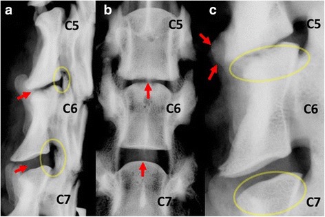
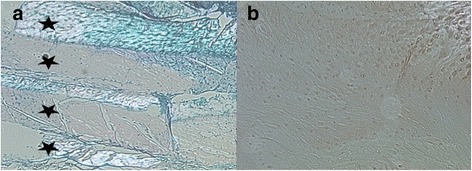
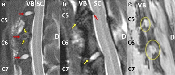
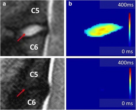
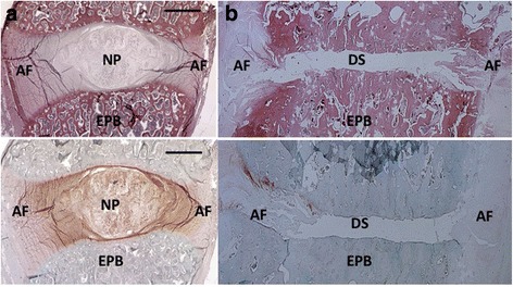
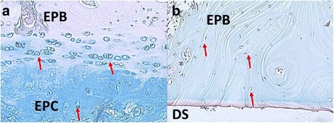
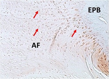
Discectomies led to a 60% drop in disc height and 24% drop in foraminal height. Segments did not fuse but showed osteophyte formation as well as endplate sclerosis. MR imaging revealed terminal degenerative changes with collapse of the disc space and loss of T2 signal intensity. The endplates showed degenerative type II Modic changes. Quantitative MR imaging revealed that over 95% of Nucleus Pulposus tissue was extracted and that the nuclear as well as overall disc hydration significantly decreased. Histology confirmed terminal degenerative changes with loss of NP tissue, loss of Annulus Fibrosus organization and loss of cartilage endplate tissue. The bony endplate displayed sclerotic changes.
Discectomies lead to terminal degenerative changes. Therefore, these procedures should be indicated with caution specifically when performed for prophylactic purposes.
椎间盘切除术是犬类脊柱椎间盘突出症常见的手术治疗方法。然而,这些手术对椎间盘组织的影响尚未完全了解。本研究的目的是在体内评估接受椎间盘切除手术的颈椎节段的退变变化。
椎间盘切除术导致椎间盘高度下降60%,椎间孔高度下降24%。节段未融合,但出现骨赘形成以及终板硬化。磁共振成像显示椎间盘间隙塌陷和T2信号强度丧失的终末期退变变化。终板显示II型Modic退变改变。定量磁共振成像显示超过95%的髓核组织被摘除,并且髓核以及整个椎间盘的水合作用显著降低。组织学证实了终末期退变变化,包括髓核组织丧失、纤维环结构丧失和软骨终板组织丧失。骨终板显示硬化改变。
椎间盘切除术会导致终末期退变变化。因此,特别是在出于预防目的进行这些手术时,应谨慎使用。