Nagesh S V Setlur, Russ M, Ionita C N, Bednarek D, Rudin S
Toshiba Stroke and Vascular Research Center, University at Buffalo.
Proc SPIE Int Soc Opt Eng. 2017 Feb 11;10137. doi: 10.1117/12.2254390. Epub 2017 Mar 13.
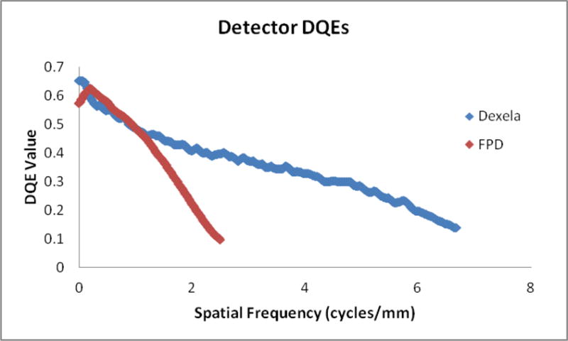
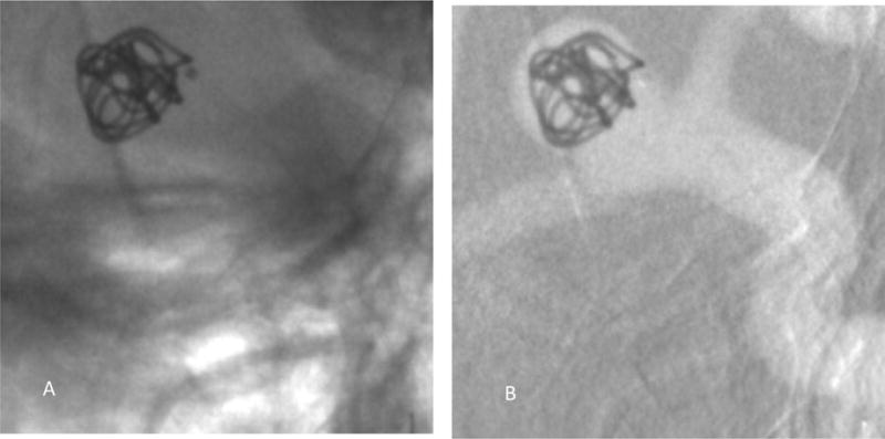
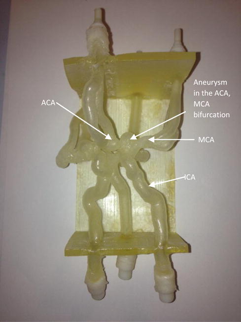
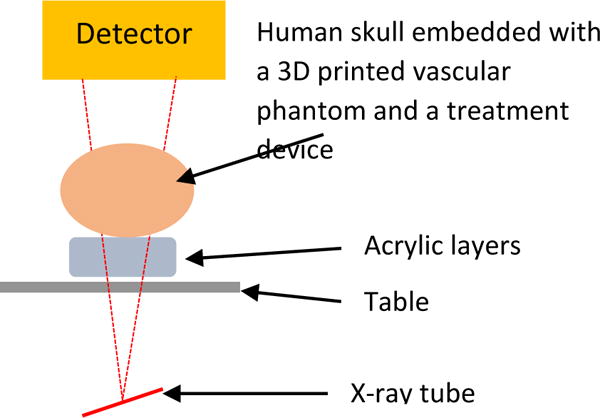
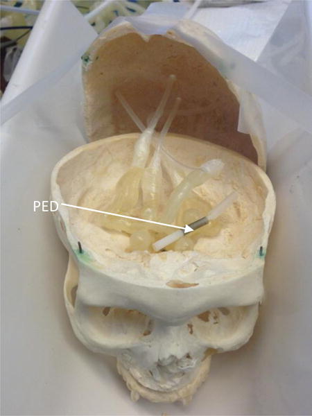
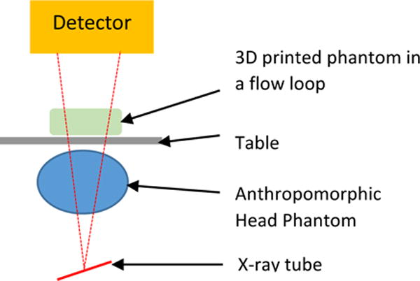
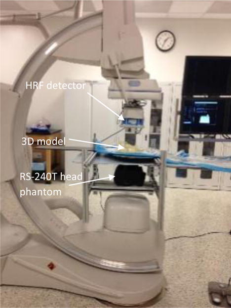
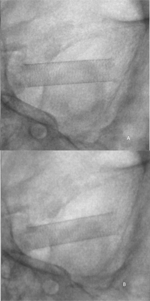
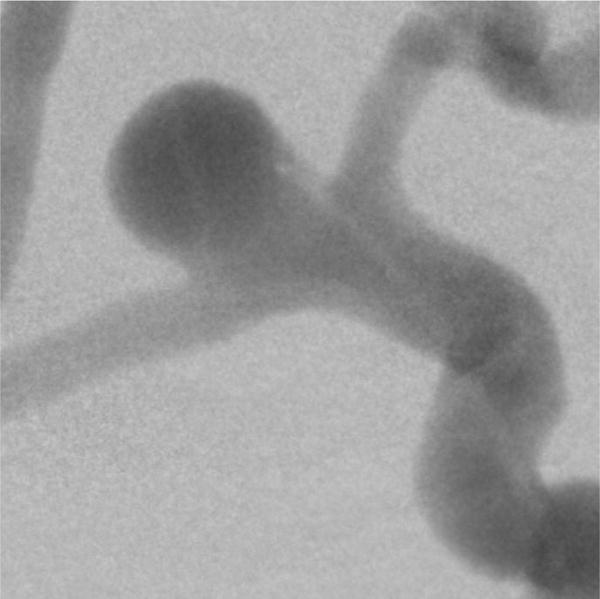
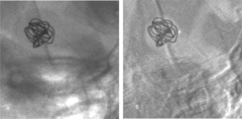
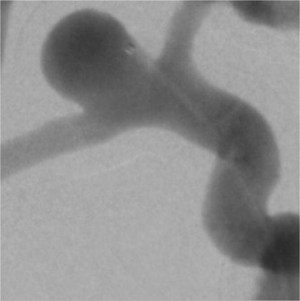
Modern 3D printing technology can fabricate vascular phantoms based on an actual human patient with a high degree of precision facilitating a realistic simulation environment for an intervention. We present two experimental setups using 3D printed patient-specific neurovasculature to simulate different disease anatomies. To simulate the human neurovasculature in the Circle of Willis, patient-based phantoms with aneurysms were 3D printed using a Objet Eden 260V printer. Anthropomorphic head phantoms and a human skull combined with acrylic plates simulated human head bone anatomy and x-ray attenuation. For dynamic studies the 3D printed phantom was connected to a pulsatile flow loop with the anthropomorphic phantom underneath. By combining different 3D printed phantoms and the anthropomorphic phantoms, different patient pathologies can be simulated. For static studies a 3D printed neurovascular phantom was embedded inside a human skull and used as a positional reference for treatment devices such as stents. To simulate tissue attenuation acrylic layers were added. Different combinations can simulate different patient treatment procedures. The Complementary-Metal-Oxide-Semiconductor (CMOS) based High Resolution Fluoroscope (HRF) with 75μm pixels offers an advantage over the state-of-the-art 200 μm pixel Flat Panel Detector (FPD) due to higher Nyquist frequency and better DQE performance. Whether this advantage is clinically useful during an actual clinical neurovascular intervention can be addressed by qualitatively evaluating images from a cohort of various cases performed using both detectors. The above-mentioned method can offer a realistic substitute for an actual clinical procedure. Also a large cohort of cases can be generated and used for a HRF clinical utility determination study.
现代3D打印技术能够基于真实人类患者高精度地制造血管模型,为介入操作营造逼真的模拟环境。我们展示了两种使用3D打印的患者特异性神经血管模型来模拟不同疾病解剖结构的实验装置。为了模拟 Willis 环中的人类神经血管,使用 Objet Eden 260V 打印机3D打印了带有动脉瘤的基于患者的模型。拟人化头部模型以及与丙烯酸板结合的人类头骨模拟了人类头部骨骼解剖结构和X射线衰减。对于动态研究,将3D打印的模型连接到脉动血流回路,其下方是拟人化模型。通过组合不同的3D打印模型和拟人化模型,可以模拟不同的患者病理情况。对于静态研究,将3D打印的神经血管模型嵌入人类头骨内,并用作支架等治疗设备的位置参考。为了模拟组织衰减,添加了丙烯酸层。不同的组合可以模拟不同的患者治疗程序。基于互补金属氧化物半导体(CMOS)的具有75μm像素的高分辨率荧光镜(HRF)由于更高的奈奎斯特频率和更好的DQE性能,比现有技术的200μm像素平板探测器(FPD)具有优势。通过定性评估使用这两种探测器进行的各种病例队列的图像,可以确定这种优势在实际临床神经血管介入过程中是否具有临床实用性。上述方法可以为实际临床操作提供逼真的替代方案。此外,可以生成大量病例队列并用于HRF临床效用确定研究。