Ma Xiaoya, Pham Vinh T, Mori Hiroyuki, MacDougald Ormond A, Shah Yatrik M, Bodary Peter F
School of Kinesiology, University of Michigan, 1402 Washington Hts., Ann Arbor, MI, United States of America.
Department of Molecular & Integrative Physiology, Ann Arbor, MI, United States of America.
PLoS One. 2017 Jun 26;12(6):e0179889. doi: 10.1371/journal.pone.0179889. eCollection 2017.
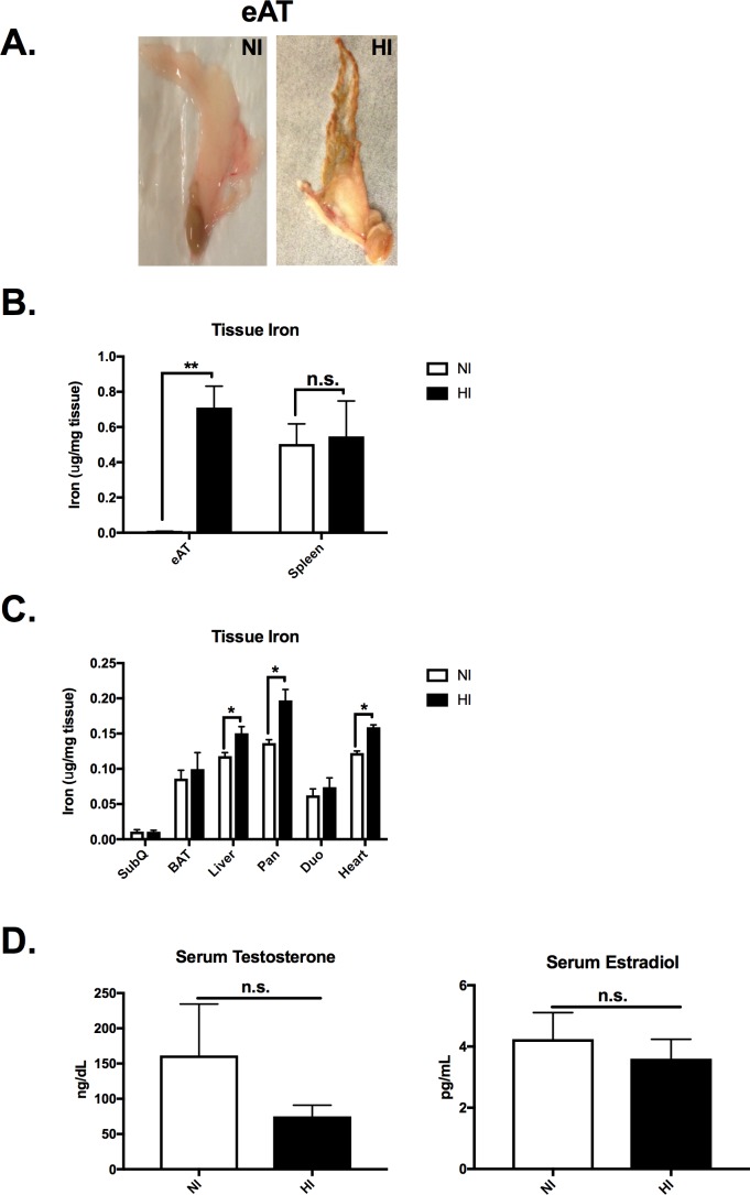
Iron dysregulation is a potential contributor to the pathology of obesity-related metabolic complications. KK/HIJ (KK) mice, a polygenic obese mouse model, have elevated serum iron levels. A subset of KK male mice display a bronzing of epididymal adipose tissue (eAT) associated with >100-fold (p<0.001) higher iron concentration.
To further phenotype and characterize the adipose tissue iron overload, 27 male KK mice were evaluated. 14 had bronzing eAT and 13 had normal appearing eAT. Fasting serum and tissues were collected for iron content, qPCR, histology and western blot.
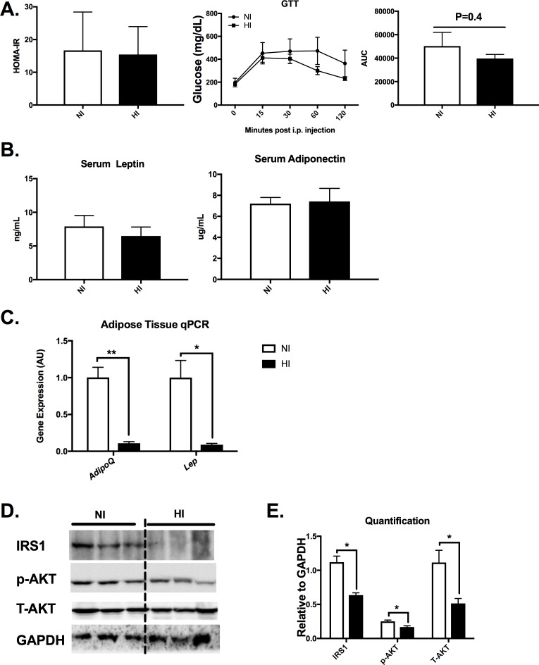
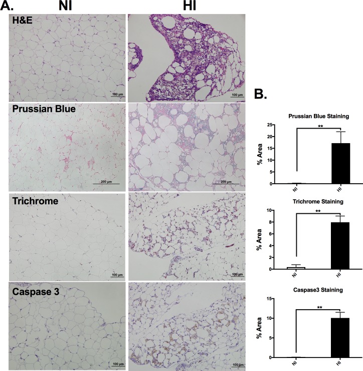
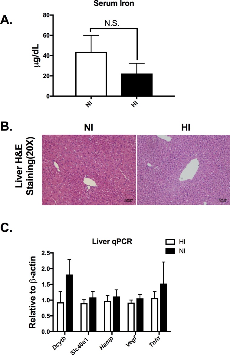
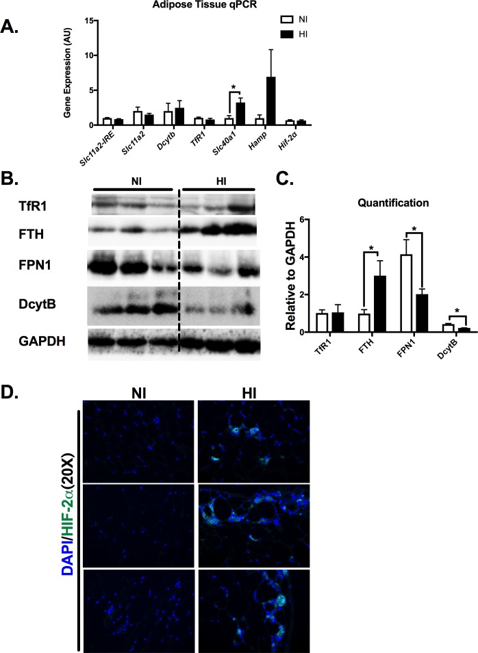
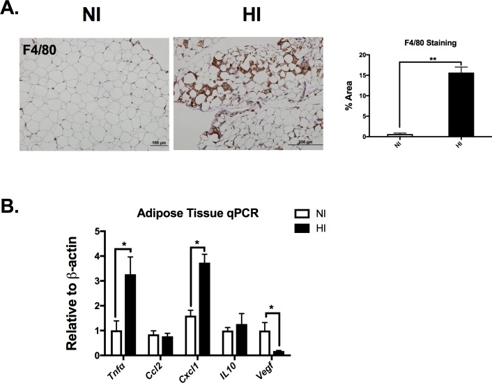
High iron levels were confirmed in bronzing eAT (High Iron group, HI) versus normal iron level (NI) in normal appearing eAT. Surprisingly, iron levels in subcutaneous and brown adipose depots were not different between the groups (p>0.05). The eAT histology revealed iron retention, macrophage clustering, tissue fibrosis, cell death as well as accumulation of HIF-2α in the high iron eAT. qPCR showed significantly decreased Lep (leptin) and AdipoQ (adiponectin), whereas Tnfα (tumor necrosis factor α), and Slc40a1 (ferroportin) were up-regulated in HI (p<0.05). Elevated HIF-2α, oxidative stress and local insulin signaling loss was also observed.
Our data suggest that deposition of iron in adipose tissue is limited to the epididymal depot in male KK mice. A robust adipose tissue remodeling is concomitant with the high iron concentration, which causes local adipose tissue insulin resistance.
铁代谢失调可能是肥胖相关代谢并发症病理过程的一个潜在因素。KK/HIJ(KK)小鼠是一种多基因肥胖小鼠模型,其血清铁水平升高。一部分KK雄性小鼠的附睾脂肪组织(eAT)出现青铜色,铁浓度升高超过100倍(p<0.001)。
为了进一步对脂肪组织铁过载进行表型分析和特征描述,对27只雄性KK小鼠进行了评估。14只小鼠的eAT出现青铜色,13只小鼠的eAT外观正常。采集空腹血清和组织用于检测铁含量、定量聚合酶链反应(qPCR)、组织学和蛋白质免疫印迹分析。
与外观正常的eAT中铁水平正常(NI)相比,出现青铜色的eAT中铁水平较高(高铁组,HI)得到证实。令人惊讶的是,两组之间皮下和棕色脂肪库中的铁水平没有差异(p>0.05)。eAT组织学显示高铁eAT中有铁潴留、巨噬细胞聚集、组织纤维化、细胞死亡以及缺氧诱导因子-2α(HIF-2α)的积累。qPCR显示在HI组中瘦素(Lep)和脂联素(AdipoQ)显著降低,而肿瘤坏死因子α(Tnfα)和铁转运蛋白1(Slc40a1)上调(p<0.05)。还观察到HIF-2α升高、氧化应激和局部胰岛素信号传导缺失。
我们的数据表明,雄性KK小鼠脂肪组织中的铁沉积仅限于附睾脂肪库。强大的脂肪组织重塑与高铁浓度同时发生,这会导致局部脂肪组织胰岛素抵抗。