• 文献检索
  • 文档翻译
  • 深度研究
  • 学术资讯
  • Suppr Zotero 插件Zotero 插件
  • 邀请有礼
  • 套餐&价格
  • 历史记录
应用&插件
Suppr Zotero 插件Zotero 插件浏览器插件Mac 客户端Windows 客户端微信小程序
定价
高级版会员购买积分包购买API积分包
服务
文献检索文档翻译深度研究API 文档MCP 服务
关于我们
关于 Suppr公司介绍联系我们用户协议隐私条款
关注我们

Suppr 超能文献

核心技术专利:CN118964589B侵权必究
粤ICP备2023148730 号-1Suppr @ 2026

文献检索

告别复杂PubMed语法,用中文像聊天一样搜索,搜遍4000万医学文献。AI智能推荐,让科研检索更轻松。

立即免费搜索

文件翻译

保留排版,准确专业,支持PDF/Word/PPT等文件格式,支持 12+语言互译。

免费翻译文档

深度研究

AI帮你快速写综述,25分钟生成高质量综述,智能提取关键信息,辅助科研写作。

立即免费体验

尸检、数字减影血管造影和计算机断层静脉造影下Labbé静脉的解剖学比较。

Anatomic comparison of veins of Labbé between autopsy, digital subtraction angiography and computed tomographic venography.

作者信息

Fang Qiong, Jiang Anhong, Tao Wei, Xin Lin

机构信息

Department of Anatomy, Anhui Medical College, Hefei, 230601, Anhui, China.

Department of Radiology, The Second Affiliated Hospital of Anhui Medical University, Hefei, 230601, China.

出版信息

Biomed Eng Online. 2017 Jun 26;16(1):84. doi: 10.1186/s12938-017-0374-3.

DOI:10.1186/s12938-017-0374-3
PMID:28651592
原文链接:https://pmc.ncbi.nlm.nih.gov/articles/PMC5485712/
Abstract

OBJECTIVE

The drainage portion of the vein of Labbé varies, and it is difficult to predict whether the operation is likely to damage this vein. The aim of this study was to correlate the microanatomy of the vein of Labbé with digital subtraction angiography (DSA) and computed tomographic venography (CTV), in order to provide a basis for the preservation of the vein of Labbé during a supratentorial surgical approach.

METHODS

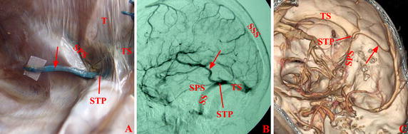
A total of 30 human cadavers (60 sides) and 61 living patients (110 sides) were examined in this study. Each cadaver head was injected with blue latex via the superior sagittal sinus and the internal jugular veins. The venograms of each patient were obtained from the venous phases of DSA (60 sides for 36 patients) or CTV (50 sides for 25 patients).

RESULTS

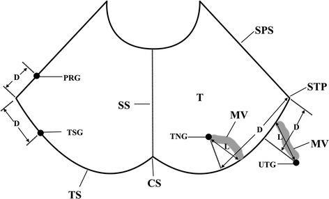
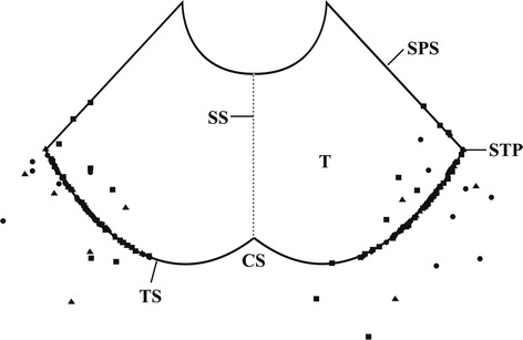
The patients were divided into four subgroups based on the location where a vein entered the dural sinus: the transverse sinus group, the tentorial group, the petrosal group, and the upper-transverse sinus group. The veins of Labbé in transverse sinus group and petrosal group directly entered dural sinus. The veins of Labbé in tentorial group and upper-transverse sinus group indirectly entered transverse sinus via the tentorium sinus or the upper-transverse sinus. These sinuses were meningeal veins running through two layers of the cerebral dura mater. The length of meningeal veins in these groups was 10.0 ± 7.2 mm. The veins of Labbé were mainly localized around the STP junction, which was the confluence of sigmoid sinus, transverse sinus, and superior petrosal sinus. The distance between the dural entrance of veins and the STP junction was 16.8 ± 10.2 mm. There was no significant difference in the results of the DSA and CTV examinations when compared to the observations in cadavers.

CONCLUSIONS

Preoperative venograms are useful to design an individualized surgical approach for the preservation of the vein of Labbé. In general, the supratentorial median approach has the least chance to damage this vein. However, when preoperative venograms show that the vein of Labbé is too close to the confluence of sinuses or the meningeal veins are too long, an alternative approach should be chosen.

摘要

目的

Labbe静脉的引流部分存在变异,难以预测手术是否可能损伤该静脉。本研究的目的是将Labbe静脉的微观解剖结构与数字减影血管造影(DSA)和计算机断层静脉造影(CTV)相关联,以便为幕上手术入路期间保留Labbe静脉提供依据。

方法

本研究共检查了30具人类尸体(60侧)和61例活体患者(110侧)。每具尸体头部通过上矢状窦和颈内静脉注入蓝色乳胶。每位患者的静脉造影图像来自DSA的静脉期(36例患者60侧)或CTV(25例患者50侧)。

结果

根据静脉进入硬脑膜窦的位置,将患者分为四个亚组:横窦组、小脑幕组、岩骨组和上横窦组。横窦组和岩骨组的Labbe静脉直接进入硬脑膜窦。小脑幕组和上横窦组的Labbe静脉通过小脑幕窦或上横窦间接进入横窦。这些窦是穿过硬脑膜两层的脑膜静脉。这些组中脑膜静脉的长度为10.0±7.2mm。Labbe静脉主要位于乙状窦、横窦和岩上窦汇合处的STP交界处周围。静脉的硬脑膜入口与STP交界处之间的距离为16.8±10.2mm。与尸体观察结果相比,DSA和CTV检查结果无显著差异。

结论

术前静脉造影有助于设计个性化的手术入路以保留Labbe静脉。一般来说,幕上正中入路损伤该静脉的机会最小。然而,当术前静脉造影显示Labbe静脉离窦汇合处太近或脑膜静脉太长时,应选择其他入路。

https://cdn.ncbi.nlm.nih.gov/pmc/blobs/2bdf/5485712/fbf0f4217657/12938_2017_374_Fig6_HTML.jpg
https://cdn.ncbi.nlm.nih.gov/pmc/blobs/2bdf/5485712/a1ccae471de1/12938_2017_374_Fig1_HTML.jpg
https://cdn.ncbi.nlm.nih.gov/pmc/blobs/2bdf/5485712/4fa28173a9a3/12938_2017_374_Fig2_HTML.jpg
https://cdn.ncbi.nlm.nih.gov/pmc/blobs/2bdf/5485712/14239c0a447c/12938_2017_374_Fig3_HTML.jpg
https://cdn.ncbi.nlm.nih.gov/pmc/blobs/2bdf/5485712/d9899160f1f1/12938_2017_374_Fig4_HTML.jpg
https://cdn.ncbi.nlm.nih.gov/pmc/blobs/2bdf/5485712/3f94b219bfb3/12938_2017_374_Fig5_HTML.jpg
https://cdn.ncbi.nlm.nih.gov/pmc/blobs/2bdf/5485712/fbf0f4217657/12938_2017_374_Fig6_HTML.jpg

相似文献

1
Anatomic comparison of veins of Labbé between autopsy, digital subtraction angiography and computed tomographic venography.尸检、数字减影血管造影和计算机断层静脉造影下Labbé静脉的解剖学比较。
Biomed Eng Online. 2017 Jun 26;16(1):84. doi: 10.1186/s12938-017-0374-3.
2
Basal vein in the posterior incisural space: An anatomical comparison based on autopsy, digital subtraction angiography and computed tomographic venography.后切迹间隙内的基底静脉:基于尸检、数字减影血管造影和计算机断层静脉造影的解剖学比较
J Xray Sci Technol. 2016;24(2):319-27. doi: 10.3233/XST-160555.
3
Utility of dynamic computed tomography angiography in the preoperative evaluation of skull base tumors.动态计算机断层血管造影在颅底肿瘤术前评估中的应用价值。
J Neurosurg. 2015 Jul;123(1):1-8. doi: 10.3171/2014.10.JNS141055. Epub 2015 Apr 3.
4
Dural entrance of the bridging vein into the transverse sinus provides a reliable measure for preoperative planning: an anatomic comparison between cadavers and neuroimages.桥静脉进入横窦的硬脑膜入口为术前规划提供了可靠的测量方法:尸体与神经影像的解剖学比较
Neurosurgery. 2008 May;62(5 Suppl 2):ONS289-95; discussion ONS295-6. doi: 10.1227/01.neu.0000326009.46197.ab.
5
Reconstruction of vein of Labbé in temporo-occipital meningioma invading transverse sinus: technical report.Labbé 静脉在侵犯横窦的颞枕部脑膜瘤中的重建:技术报告。
Acta Neurochir (Wien). 2010 Jun;152(6):941-5; discussion 945. doi: 10.1007/s00701-009-0572-2. Epub 2009 Dec 15.
6
Analysis of Temporobasal Vein with Short Subdural Segment for Anterior Transpetrosal Approach.分析颞底静脉及其短硬脑膜下段在前经岩骨入路中的作用。
World Neurosurg. 2019 Dec;132:e554-e562. doi: 10.1016/j.wneu.2019.08.077. Epub 2019 Aug 21.
7
The dural entrance of cerebral bridging veins into the superior sagittal sinus: an anatomical comparison between cadavers and digital subtraction angiography.大脑桥静脉进入上矢状窦的硬脑膜入口:尸体与数字减影血管造影的解剖学比较
Neuroradiology. 2007 Feb;49(2):169-75. doi: 10.1007/s00234-006-0175-z. Epub 2007 Jan 3.
8
[Detection of the venous system of the skull base using three-dimensional CT angiography (3D-CTA): utility of the subtemporal approach].[使用三维CT血管造影术(3D-CTA)检测颅底静脉系统:颞下入路的实用性]
No Shinkei Geka. 2000 Jan;28(1):17-22.
9
Preoperative evaluation of venous systems with 3-dimensional contrast-enhanced magnetic resonance venography in brain tumors: comparison with time-of-flight magnetic resonance venography and digital subtraction angiography.三维对比增强磁共振静脉血管造影术在脑肿瘤静脉系统术前评估中的应用:与时间飞跃磁共振静脉血管造影术及数字减影血管造影术的比较
Surg Neurol. 2005 Aug;64(2):128-33; discussion 133-4. doi: 10.1016/j.surneu.2004.10.029.
10
Anatomical studies on the temporal bridging veins with Dextroscope and its application in tumor surgery across the middle and posterior fossa.使用Dextroscope对颞部桥静脉进行解剖学研究及其在中后颅窝肿瘤手术中的应用
Clin Neurol Neurosurg. 2011 Dec;113(10):889-94. doi: 10.1016/j.clineuro.2011.06.008. Epub 2011 Aug 9.

引用本文的文献

1
Magnetic resonance venography study of venous structures of the transverse-sigmoid sinus region in the Han Chinese population: A cross-sectional study.汉族人群横窦-乙状窦区域静脉结构的磁共振静脉成像研究:一项横断面研究。
SAGE Open Med. 2024 Oct 29;12:20503121241274244. doi: 10.1177/20503121241274244. eCollection 2024.
2
Simulation of the occipital transtentorial approach incorporating visualization of the cerebellar tentorium using three-dimensional computed tomography angiography and gadolinium-enhanced T1-weighted magnetic resonance imaging: technical note.应用三维 CT 血管造影和钆增强 T1 加权磁共振成像对小脑幕进行可视化处理的枕下入路模拟:技术说明。
Neurosurg Rev. 2023 Sep 29;46(1):259. doi: 10.1007/s10143-023-02170-6.
3

本文引用的文献

1
Computed tomographic angiography of the superficial cerebral venous anastomosis based on volume rendering, multi-planar reconstruction, and integral imaging display.基于容积再现、多平面重建和整体成像显示的大脑浅静脉吻合的计算机断层血管造影。
Australas Phys Eng Sci Med. 2015 Dec;38(4):777-83. doi: 10.1007/s13246-015-0403-0. Epub 2015 Nov 17.
2
Postoperative cerebral venous infarction.术后脑静脉梗死
J Pediatr Neurosci. 2015 Jan-Mar;10(1):5-8. doi: 10.4103/1817-1745.154314.
3
What is the risk of venous infarction to intra-operative sacrifice of either the superficial or deep cerebral bridging veins?
A New Classification of the Anatomical Variations of Labbé's Inferior Anastomotic Vein.拉贝下吻合静脉解剖变异的新分类。
Tomography. 2022 Aug 30;8(5):2182-2192. doi: 10.3390/tomography8050183.
4
Cortical Vein Thrombosis after Infliximab Treatment for Crohn's Disease.英夫利昔单抗治疗克罗恩病后发生的皮质静脉血栓形成
Neurol Int. 2021 Mar 11;13(1):120-124. doi: 10.3390/neurolint13010011.
术中牺牲大脑浅或深桥静脉时,静脉梗死的风险有多大?
Childs Nerv Syst. 2014 May;30(5):811-3. doi: 10.1007/s00381-014-2405-8. Epub 2014 Mar 27.
4
Anatomical variations of the vein of Labbé: an angiographic study.Labbe静脉的解剖变异:一项血管造影研究。
Surg Radiol Anat. 2014 Oct;36(8):769-73. doi: 10.1007/s00276-014-1264-z. Epub 2014 Feb 15.
5
Cerebral venous infarction: a potentially avoidable complication of deep brain stimulation surgery.脑静脉梗死:深部脑刺激手术一种潜在可避免的并发症。
Neuromodulation. 2013 Sep-Oct;16(5):407-13; discussion 413. doi: 10.1111/ner.12052. Epub 2013 Jun 5.
6
Post-ischemic administration of vascular endothelial growth factor inhibitor in a rat model of cerebral venous infarction.在大鼠脑静脉梗死模型中缺血后给予血管内皮生长因子抑制剂
Neurol Med Chir (Tokyo). 2013;53(3):135-40. doi: 10.2176/nmc.53.135.
7
The intracranial bridging veins: a comprehensive review of their history, anatomy, histology, pathology, and neurosurgical implications.颅内桥静脉:对其历史、解剖学、组织学、病理学及神经外科意义的全面综述
Childs Nerv Syst. 2013 Jul;29(7):1073-8. doi: 10.1007/s00381-013-2054-3. Epub 2013 Mar 2.
8
External landmarks for identifying the drainage site of the vein of Labbé: application to neurosurgical procedures.用于确定Labbe静脉引流部位的体表标志:在神经外科手术中的应用。
Br J Neurosurg. 2012 Jun;26(3):383-5. doi: 10.3109/02688697.2011.631620. Epub 2011 Nov 22.
9
Contemporary angiographic assessment and clinical implications of the vein of labbé in neurotologic surgery.神经耳科学手术中 Labbe 静脉的当代血管造影评估及其临床意义。
Otol Neurotol. 2011 Aug;32(6):1012-6. doi: 10.1097/MAO.0b013e3182255980.
10
Incidence of venous infarction after sacrificing middle-third superior sagittal sinus cortical bridging veins in a pediatric population.小儿人群中牺牲上矢状窦中三分之一皮质桥静脉后静脉梗死的发生率。
J Neurosurg Pediatr. 2011 Mar;7(3):224-8. doi: 10.3171/2010.11.PEDS09261.