• 文献检索
  • 文档翻译
  • 深度研究
  • 学术资讯
  • Suppr Zotero 插件Zotero 插件
  • 邀请有礼
  • 套餐&价格
  • 历史记录
应用&插件
Suppr Zotero 插件Zotero 插件浏览器插件Mac 客户端Windows 客户端微信小程序
定价
高级版会员购买积分包购买API积分包
服务
文献检索文档翻译深度研究API 文档MCP 服务
关于我们
关于 Suppr公司介绍联系我们用户协议隐私条款
关注我们

Suppr 超能文献

核心技术专利:CN118964589B侵权必究
粤ICP备2023148730 号-1Suppr @ 2026

文献检索

告别复杂PubMed语法,用中文像聊天一样搜索,搜遍4000万医学文献。AI智能推荐,让科研检索更轻松。

立即免费搜索

文件翻译

保留排版,准确专业,支持PDF/Word/PPT等文件格式,支持 12+语言互译。

免费翻译文档

深度研究

AI帮你快速写综述,25分钟生成高质量综述,智能提取关键信息,辅助科研写作。

立即免费体验

使用高分辨率磁共振成像对发育性遗忘症内侧颞叶亚区进行容积分析。

Volumetric analysis of medial temporal lobe subregions in developmental amnesia using high-resolution magnetic resonance imaging.

机构信息

Rotman Research Institute at Baycrest, Toronto, Ontario, Canada.

出版信息

Hippocampus. 2013 Oct;23(10):855-60. doi: 10.1002/hipo.22153. Epub 2013 Jul 5.

DOI:10.1002/hipo.22153
PMID:23749334
原文链接:https://pmc.ncbi.nlm.nih.gov/articles/PMC4165307/
Abstract

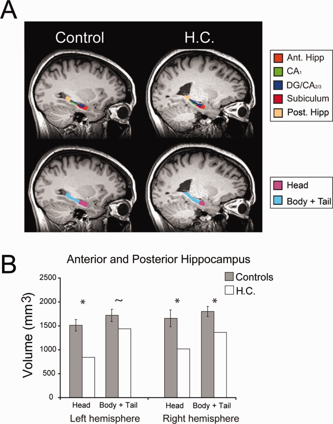
There is great interest in the cognitive consequences of hippocampal volume loss in developmental amnesia (DA). In many DA cases, volume loss occurs before the hippocampus is fully developed, and yet little is known about the locus, extent, and distribution of damage in these cases. We used high-resolution MRI to manually segment the medial temporal lobe (MTL) subregions in H.C., an adult with DA, and a group of sex-, age- and education-matched control participants (n = 10). The hippocampus was defined and divided into anterior (head) and posterior (body and tail) segments. Within the body of the hippocampus, the subregions (CA1 , DG/CA2/3 , and subiculum) were defined. Finally, the entorhinal (ERC), perirhinal (PRC), and parahippocampal (PHC) cortices were segmented. Anterior hippocampus was reduced bilaterally and posterior hippocampus was significantly reduced on the right. In the body of the hippocampus, all three subregions were reduced in the left hemisphere, whereas CA1 and subiculum were reduced in the right hemisphere. No group differences were observed in the PRC and ERC, whereas left PHC volume was marginally increased in H.C. compared to controls. These results can be used to inform patterns of spared and impaired cognitive abilities in DA and perhaps in amnesia more generally.

摘要

人们对发育性遗忘症 (DA) 中海马体积损失的认知后果非常感兴趣。在许多 DA 病例中,海马体积的损失发生在海马完全发育之前,但对于这些病例中的损伤部位、程度和分布,人们知之甚少。我们使用高分辨率 MRI 手动分割了 H.C.(一名患有 DA 的成年人)和一组性别、年龄和教育程度匹配的对照组参与者(n = 10)的内侧颞叶 (MTL) 亚区。定义并将海马体分为前(头)部和后(体和尾)部两个部分。在海马体的主体内,定义了亚区(CA1、DG/CA2/3 和 subiculum)。最后,分割了内嗅皮质 (ERC)、旁嗅皮质 (PRC) 和海马旁回皮质 (PHC)。双侧前海马体减少,右侧后海马体显著减少。在海马体的主体内,左侧的所有三个亚区都减少了,而右侧的 CA1 和 subiculum 也减少了。在 PRC 和 ERC 中没有观察到组间差异,而与对照组相比,H.C. 的左侧 PHC 体积略有增加。这些结果可用于告知 DA 中的认知能力保留和受损模式,也许更广泛地说,在遗忘症中也是如此。

https://cdn.ncbi.nlm.nih.gov/pmc/blobs/ecdc/4165307/4059b6f08e2c/hipo0023-0855-f2.jpg
https://cdn.ncbi.nlm.nih.gov/pmc/blobs/ecdc/4165307/9604cc3015b6/hipo0023-0855-f1.jpg
https://cdn.ncbi.nlm.nih.gov/pmc/blobs/ecdc/4165307/4059b6f08e2c/hipo0023-0855-f2.jpg
https://cdn.ncbi.nlm.nih.gov/pmc/blobs/ecdc/4165307/9604cc3015b6/hipo0023-0855-f1.jpg
https://cdn.ncbi.nlm.nih.gov/pmc/blobs/ecdc/4165307/4059b6f08e2c/hipo0023-0855-f2.jpg

相似文献

1
Volumetric analysis of medial temporal lobe subregions in developmental amnesia using high-resolution magnetic resonance imaging.使用高分辨率磁共振成像对发育性遗忘症内侧颞叶亚区进行容积分析。
Hippocampus. 2013 Oct;23(10):855-60. doi: 10.1002/hipo.22153. Epub 2013 Jul 5.
2
A protocol for manual segmentation of medial temporal lobe subregions in 7 Tesla MRI.一种在7特斯拉磁共振成像中手动分割内侧颞叶亚区域的方案。
Neuroimage Clin. 2017 May 26;15:466-482. doi: 10.1016/j.nicl.2017.05.022. eCollection 2017.
3
Mesial temporal damage in temporal lobe epilepsy: a volumetric MRI study of the hippocampus, amygdala and parahippocampal region.颞叶癫痫中的内侧颞叶损伤:海马体、杏仁核及海马旁区域的容积磁共振成像研究
Brain. 2003 Feb;126(Pt 2):462-9. doi: 10.1093/brain/awg034.
4
Downstream effects of polypathology on neurodegeneration of medial temporal lobe subregions.多病理学对内侧颞叶亚区神经退行性变的下游影响。
Acta Neuropathol Commun. 2021 Jul 21;9(1):128. doi: 10.1186/s40478-021-01225-3.
5
Differential connectivity of perirhinal and parahippocampal cortices within human hippocampal subregions revealed by high-resolution functional imaging.高分辨率功能成像显示人类海马亚区中海马旁回和旁海马皮质的差异连接。
J Neurosci. 2012 May 9;32(19):6550-60. doi: 10.1523/JNEUROSCI.3711-11.2012.
6
Quantifying medial temporal lobe damage in memory-impaired patients.量化记忆受损患者的内侧颞叶损伤。
Hippocampus. 2005;15(1):79-85. doi: 10.1002/hipo.20032.
7
Quantitative comparison of 21 protocols for labeling hippocampal subfields and parahippocampal subregions in in vivo MRI: towards a harmonized segmentation protocol.21种用于在活体磁共振成像中标记海马亚区和海马旁回亚区域的方案的定量比较:迈向统一的分割方案
Neuroimage. 2015 May 1;111:526-41. doi: 10.1016/j.neuroimage.2015.01.004. Epub 2015 Jan 14.
8
Hippocampal subfield volumes at 7T in early Alzheimer's disease and normal aging.7T下早期阿尔茨海默病和正常衰老的海马亚区体积
Neurobiol Aging. 2014 Sep;35(9):2039-45. doi: 10.1016/j.neurobiolaging.2014.02.021. Epub 2014 Mar 3.
9
Age differences in hippocampal subfield volumes from childhood to late adulthood.从童年到成年晚期海马体亚区体积的年龄差异。
Hippocampus. 2016 Feb;26(2):220-8. doi: 10.1002/hipo.22517. Epub 2015 Sep 4.
10
Temporal lobe epilepsy affects spatial organization of entorhinal cortex connectivity.颞叶癫痫会影响内嗅皮层连接的空间组织。
Epilepsy Behav. 2018 Nov;88:87-95. doi: 10.1016/j.yebeh.2018.06.038. Epub 2018 Sep 20.

引用本文的文献

1
Developing an Anatomically Valid Segmentation Protocol for Anterior Regions of the Medial Temporal Lobe for Neurodegenerative Diseases.为神经退行性疾病开发一种用于内侧颞叶前部区域的解剖学上有效的分割方案。
Hippocampus. 2025 Sep;35(5):e70027. doi: 10.1002/hipo.70027.
2
Effects of Early Midlife Ovarian Removal on Medial Temporal Lobe Gray Matter Volume and Recognition Memory.中年早期卵巢切除对内侧颞叶灰质体积和识别记忆的影响。
Hippocampus. 2025 May;35(3):e70012. doi: 10.1002/hipo.70012.
3
The interactive effects of divided attention and semantic elaboration on associative recognition memory: an fMRI study.

本文引用的文献

1
Long-axis specialization of the human hippocampus.人类海马体的长轴特化。
Trends Cogn Sci. 2013 May;17(5):230-40. doi: 10.1016/j.tics.2013.03.005. Epub 2013 Apr 16.
2
Multi-voxel pattern analysis in human hippocampal subfields.多体素模式分析在人类海马亚区中的应用。
Front Hum Neurosci. 2012 Oct 18;6:290. doi: 10.3389/fnhum.2012.00290. eCollection 2012.
3
Long-term neuropsychological, neuroanatomical, and life outcome in hippocampal amnesia.海马体遗忘症的长期神经心理学、神经解剖学和生活结局。
分散注意力和语义加工对联想识别记忆的交互作用:一项功能磁共振成像研究
Cereb Cortex. 2024 Dec 3;34(12). doi: 10.1093/cercor/bhae464.
4
Developing an anatomically valid segmentation protocol for anterior regions of the medial temporal lobe for neurodegenerative diseases.为神经退行性疾病开发一种用于内侧颞叶前部区域的解剖学上有效的分割方案。
bioRxiv. 2025 Feb 13:2025.02.11.637506. doi: 10.1101/2025.02.11.637506.
5
The Effect of Segmentation Method on Medial Temporal Lobe Subregion Volumes in Aging.分割方法对衰老内侧颞叶亚区体积的影响。
Hum Brain Mapp. 2024 Oct 15;45(15):e70054. doi: 10.1002/hbm.70054.
6
Postmortem imaging reveals patterns of medial temporal lobe vulnerability to tau pathology in Alzheimer's disease.尸检后影像学显示阿尔茨海默病中海马内侧颞叶对 tau 病理学的易损性模式。
Nat Commun. 2024 Jun 5;15(1):4803. doi: 10.1038/s41467-024-49205-0.
7
Comparison of histological delineations of medial temporal lobe cortices by four independent neuroanatomy laboratories.四家独立神经解剖实验室对内侧颞叶皮质的组织学描绘比较。
Hippocampus. 2024 May;34(5):241-260. doi: 10.1002/hipo.23602. Epub 2024 Feb 28.
8
Disturbed sleep is associated with reduced verbal episodic memory and entorhinal cortex volume in younger middle-aged women with risk-reducing early ovarian removal.有卵巢切除术风险的年轻中年女性中,睡眠紊乱与言语情节记忆减退和内嗅皮层体积减少有关。
Front Endocrinol (Lausanne). 2023 Oct 3;14:1265470. doi: 10.3389/fendo.2023.1265470. eCollection 2023.
9
Ex vivo MRI atlas of the human medial temporal lobe: characterizing neurodegeneration due to tau pathology.人类内侧颞叶的离体 MRI 图谱:特征性tau 病理学所致神经退行性变。
Acta Neuropathol Commun. 2021 Oct 24;9(1):173. doi: 10.1186/s40478-021-01275-7.
10
Getting better without memory.失忆后逐渐康复。
Soc Cogn Affect Neurosci. 2020 Oct 8;15(8):815-825. doi: 10.1093/scan/nsaa105.
Clin Neuropsychol. 2012;26(2):335-69. doi: 10.1080/13854046.2012.655781. Epub 2012 Mar 8.
4
Patient HC with developmental amnesia can construct future scenarios.患者 HC 患有发展性遗忘症,可以构建未来场景。
Neuropsychologia. 2011 Nov;49(13):3620-8. doi: 10.1016/j.neuropsychologia.2011.09.015. Epub 2011 Sep 22.
5
Impaired event memory and recollection in a case of developmental amnesia.一例发育性遗忘症患者的事件记忆和回忆受损。
Neurocase. 2011 Oct;17(5):394-409. doi: 10.1080/13554794.2010.532138. Epub 2011 Jun 30.
6
Postnatal development of the hippocampal formation: a stereological study in macaque monkeys.海马结构的产后发育:猕猴的体视学研究。
J Comp Neurol. 2011 Apr 15;519(6):1051-70. doi: 10.1002/cne.22549.
7
Deficits in past remembering extend to future imagining in a case of developmental amnesia.发展性遗忘症患者的过去记忆缺陷会延伸至未来想象。
Neuropsychologia. 2010 Sep;48(11):3179-86. doi: 10.1016/j.neuropsychologia.2010.06.011. Epub 2010 Jun 16.
8
Imaging the human medial temporal lobe with high-resolution fMRI.高分辨率 fMRI 成像人类内侧颞叶。
Neuron. 2010 Feb 11;65(3):298-308. doi: 10.1016/j.neuron.2009.12.022.
9
Developing an animal model of human amnesia: the role of the hippocampus.开发人类遗忘症的动物模型:海马的作用。
Neuropsychologia. 2010 Jul;48(8):2290-302. doi: 10.1016/j.neuropsychologia.2009.10.027. Epub 2009 Oct 31.
10
Performance-related sustained and anticipatory activity in human medial temporal lobe during delayed match-to-sample.延迟样本匹配任务中人类内侧颞叶与表现相关的持续和预期活动。
J Neurosci. 2009 Sep 23;29(38):11880-90. doi: 10.1523/JNEUROSCI.2245-09.2009.