Yi Faliu, Huang Junzhou, Yang Lin, Xie Yang, Xiao Guanghua
University of Texas Southwestern Medical Center, Quantitative Biomedical Research Center, Department of Clinical Science, Dallas, Texas, United States.
University of Texas at Arlington, Department of Computer Science and Engineering, Arlington, Texas, United States.
J Med Imaging (Bellingham). 2017 Apr;4(2):027502. doi: 10.1117/1.JMI.4.2.027502. Epub 2017 Jun 21.
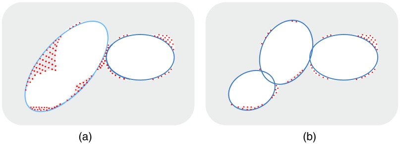
Extraction of cell nuclei from hematoxylin and eosin (H&E)-stained histopathological images is an essential preprocessing step in computerized image analysis for disease detection, diagnosis, and prognosis. We present an automated cell nuclei segmentation approach that works with H&E-stained images. A color deconvolution algorithm was first applied to the image to get the hematoxylin channel. Using a morphological operation and thresholding technique on the hematoxylin channel image, candidate target nuclei and background regions were detected, which were then used as markers for a marker-controlled watershed transform segmentation algorithm. Moreover, postprocessing was conducted to split the touching nuclei. For each segmented region from the previous steps, the regional maximum value positions were identified as potential nuclei centers. These maximum values were further grouped into [Formula: see text]-clusters, and the locations within each cluster were connected with the minimum spanning tree technique. Then, these connected positions were utilized as new markers for a watershed segmentation approach. The final number of nuclei at each region was determined by minimizing an objective function that iterated all of the possible [Formula: see text]-values. The proposed method was applied to the pathological images of the tumor tissues from The Cancer Genome Atlas study. Experimental results show that the proposed method can lead to promising results in terms of segmentation accuracy and separation of touching nuclei.
从苏木精和伊红(H&E)染色的组织病理学图像中提取细胞核是疾病检测、诊断和预后的计算机图像分析中必不可少的预处理步骤。我们提出了一种适用于H&E染色图像的自动细胞核分割方法。首先将颜色反卷积算法应用于图像以获得苏木精通道。对苏木精通道图像使用形态学操作和阈值技术,检测出候选目标细胞核和背景区域,然后将其用作标记控制分水岭变换分割算法的标记。此外,进行后处理以分割相互接触的细胞核。对于上一步骤中分割出的每个区域,将区域最大值位置识别为潜在的细胞核中心。这些最大值进一步被分组为[公式:见原文]-簇,并使用最小生成树技术连接每个簇内的位置。然后,将这些连接位置用作分水岭分割方法的新标记。通过最小化迭代所有可能的[公式:见原文]-值的目标函数来确定每个区域的最终细胞核数量。所提出的方法应用于来自癌症基因组图谱研究的肿瘤组织病理图像。实验结果表明,所提出的方法在分割精度和接触细胞核分离方面能取得有前景的结果。