Barash N R, Nosala C, Pham J K, McInally S G, Gourguechon S, McCarthy-Sinclair B, Dawson S C
Department of Microbiology and Molecular Genetics, UC Davis, Davis, California, USA.
Department of Molecular and Cell Biology, UC Berkeley, Berkeley, California, USA.
mSphere. 2017 Jun 21;2(3). doi: 10.1128/mSphere.00343-16. eCollection 2017 May-Jun.
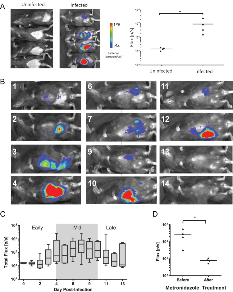
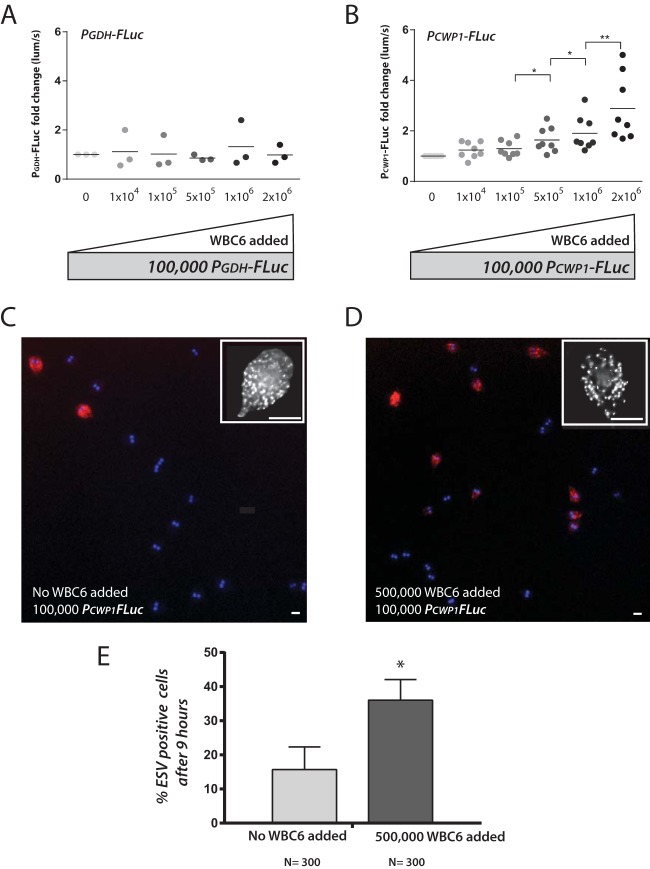
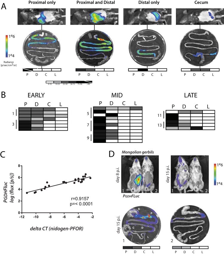
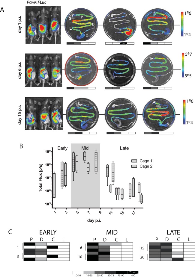
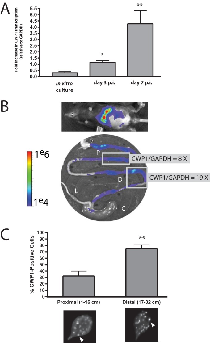
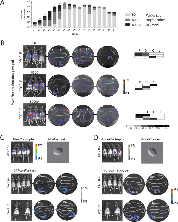
is a highly prevalent yet understudied protistan parasite causing significant diarrheal disease worldwide. Hosts ingest cysts from contaminated sources. In the gastrointestinal tract, cysts excyst to become motile trophozoites, colonizing and attaching to the gut epithelium. Trophozoites later differentiate into infectious cysts that are excreted and contaminate the environment. Due to the limited accessibility of the gut, the temporospatial dynamics of giardiasis in the host are largely inferred from laboratory culture and thus may not mirror physiology in the host. Here, we have developed bioluminescent imaging (BLI) to directly interrogate and quantify the temporospatial dynamics of infection, thereby providing an improved murine model to evaluate anti- drugs. Using BLI, we determined that parasites primarily colonize the proximal small intestine nonuniformly in high-density foci. By imaging encystation-specific bioreporters, we show that encystation initiates shortly after inoculation and continues throughout the duration of infection. Encystation also initiates in high-density foci in the proximal small intestine, and high density contributes to the initiation of encystation in laboratory culture. We suggest that these high-density foci of colonizing and encysting likely result in localized disruption to the epithelium. This more accurate visualization of giardiasis redefines the dynamics of the life cycle, paving the way for future mechanistic studies of density-dependent parasitic processes in the host. is a single-celled parasite causing significant diarrheal disease in several hundred million people worldwide. Due to limited access to the site of infection in the gastrointestinal tract, our understanding of the dynamics of infections in the host has remained limited and largely inferred from laboratory culture. To better understand physiology and colonization in the host, we developed imaging methods to quantify expressing bioluminescent physiological reporters in two relevant animal models. We discovered that parasites primarily colonize and encyst in the proximal small intestine in discrete, high-density foci. We also show that high parasite density contributes to encystation initiation.
是一种高度流行但研究不足的原生动物寄生虫,在全球范围内导致严重的腹泻疾病。宿主从受污染的来源摄入囊肿。在胃肠道中,囊肿脱囊成为活动的滋养体,定殖并附着于肠道上皮。滋养体随后分化为感染性囊肿,这些囊肿被排出并污染环境。由于肠道难以接近,贾第虫病在宿主体内的时空动态很大程度上是从实验室培养中推断出来的,因此可能无法反映宿主体内的生理学情况。在这里,我们开发了生物发光成像(BLI)技术,以直接探究和量化感染的时空动态,从而提供一个改进的小鼠模型来评估抗寄生虫药物。使用BLI,我们确定寄生虫主要在近端小肠以高密度病灶的形式非均匀定殖。通过对特定包囊化生物报告基因进行成像,我们表明包囊化在接种后不久就开始,并在整个感染期间持续进行。包囊化也在近端小肠的高密度病灶中开始,并且高密度有助于实验室培养中包囊化的起始。我们认为这些定殖和包囊化的高密度病灶可能导致上皮局部破坏。这种对贾第虫病更准确的可视化重新定义了生命周期的动态,为未来对宿主体内密度依赖性寄生过程的机制研究铺平了道路。是一种单细胞寄生虫,在全球数亿人中引起严重的腹泻疾病。由于难以接近胃肠道中的感染部位,我们对宿主体内感染动态的理解仍然有限,并且很大程度上是从实验室培养中推断出来的。为了更好地了解宿主体内的生理学和定殖情况,我们开发了成像方法,以量化在两种相关动物模型中表达生物发光生理学报告基因的情况。我们发现寄生虫主要在近端小肠以离散的高密度病灶形式定殖和包囊。我们还表明高寄生虫密度有助于包囊化的起始。