Charng Jason, Jacobson Samuel G, Heon Elise, Roman Alejandro J, McGuigan David B, Sheplock Rebecca, Kosyk Mychajlo S, Swider Malgorzata, Cideciyan Artur V
Scheie Eye Institute, Perelman School of Medicine, University of Pennsylvania, Philadelphia, Pennsylvania, United States.
Department of Ophthalmology and Vision Sciences, The Hospital for Sick Children, University of Toronto, Toronto, Ontario, Canada.
Invest Ophthalmol Vis Sci. 2017 Jun 1;58(7):3215-3224. doi: 10.1167/iovs.17-21909.
Pupillary light reflex (PLR) is driven by outer retinal photoreceptors and by melanopsin-expressing intrinsically photosensitive retinal ganglion cells of the inner retina. To isolate the melanopic component, we studied patients with severe vision loss due to Leber congenital amaurosis (LCA) caused by gene mutations acting on the outer retina.
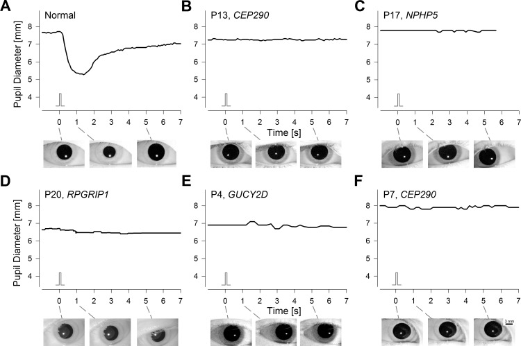
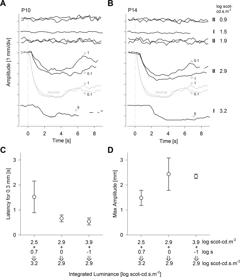
Direct PLR was recorded in LCA patients (n = 21) with known molecular causation and severe vision loss. Standard stimuli (2.5 log scot-cd.m-2; ∼13 log quanta.cm-2.s-1; achromatic full-field) with 0.1- or 5-second duration were used in all patients. Additional recordings were performed with higher luminance (3.9 log scot-cd.m-2) in a subset of patients.
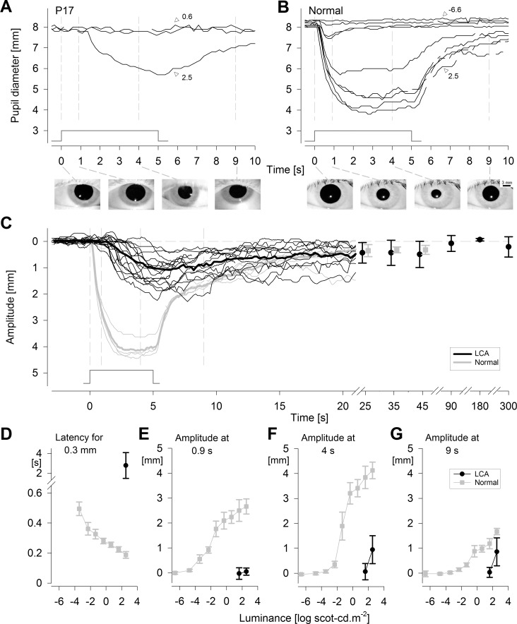
The LCA patients showed no detectable PLR to the standard stimulus with short duration. With longer-duration stimuli, a PLR was detectable in the majority (18/21) of patients. The latency of the PLR was 2.8 ± 1.3 seconds, whereas normal latency was 0.19 ± 0.02 seconds. Peak contraction amplitude in patients was 1.1 ± 0.9 mm at 6.2 ± 2.3 seconds, considerably different from normal amplitude of 4.2 ± 0.4 mm at 3.0 ± 0.4 seconds. Recordings with higher luminance demonstrated that PLRs in severe LCA could also be evoked with short-duration stimuli.
The PLR in severe LCA patients likely represents the activation of the melanopic circuit in isolation from rod and cone input. Knowledge of the properties of the human melanopic PLR allows not only comparison to those in animal models but also serves to define the fidelity of postretinal transmission in clinical trials targeting patients with no outer retinal function.
瞳孔光反射(PLR)由视网膜外层光感受器以及视网膜内层表达黑视蛋白的内在光敏视网膜神经节细胞驱动。为了分离黑视蛋白成分,我们研究了因作用于视网膜外层的基因突变导致莱伯先天性黑蒙(LCA)而视力严重丧失的患者。
记录了21例已知分子病因且视力严重丧失的LCA患者的直接PLR。所有患者均使用持续时间为0.1秒或5秒的标准刺激(2.5 log scot-cd·m⁻²;约13 log量子·cm⁻²·s⁻¹;消色差全视野)。部分患者还使用更高亮度(3.9 log scot-cd·m⁻²)进行了额外记录。
LCA患者对短持续时间的标准刺激未检测到可察觉的PLR。对于持续时间较长的刺激,大多数(18/21)患者可检测到PLR。PLR的潜伏期为2.8±1.3秒,而正常潜伏期为0.19±0.02秒。患者的峰值收缩幅度在6.2±2.3秒时为1.1±0.9毫米,与正常幅度在3.0±0.4秒时的4.2±0.4毫米有显著差异。更高亮度的记录表明,严重LCA患者的PLR也可由短持续时间刺激诱发。
严重LCA患者的PLR可能代表了黑视蛋白通路在与视杆和视锥输入分离的情况下的激活。了解人类黑视蛋白PLR的特性不仅有助于与动物模型进行比较,还可用于在针对无视网膜外层功能患者的临床试验中定义视网膜后传递的保真度。