• 文献检索
  • 文档翻译
  • 深度研究
  • 学术资讯
  • Suppr Zotero 插件Zotero 插件
  • 邀请有礼
  • 套餐&价格
  • 历史记录
应用&插件
Suppr Zotero 插件Zotero 插件浏览器插件Mac 客户端Windows 客户端微信小程序
定价
高级版会员购买积分包购买API积分包
服务
文献检索文档翻译深度研究API 文档MCP 服务
关于我们
关于 Suppr公司介绍联系我们用户协议隐私条款
关注我们

Suppr 超能文献

核心技术专利:CN118964589B侵权必究
粤ICP备2023148730 号-1Suppr @ 2026

文献检索

告别复杂PubMed语法,用中文像聊天一样搜索,搜遍4000万医学文献。AI智能推荐,让科研检索更轻松。

立即免费搜索

文件翻译

保留排版,准确专业,支持PDF/Word/PPT等文件格式,支持 12+语言互译。

免费翻译文档

深度研究

AI帮你快速写综述,25分钟生成高质量综述,智能提取关键信息,辅助科研写作。

立即免费体验

视觉回路利用互补机制来支持瞬态和持续性瞳孔收缩。

A visual circuit uses complementary mechanisms to support transient and sustained pupil constriction.

作者信息

Keenan William Thomas, Rupp Alan C, Ross Rachel A, Somasundaram Preethi, Hiriyanna Suja, Wu Zhijian, Badea Tudor C, Robinson Phyllis R, Lowell Bradford B, Hattar Samer S

机构信息

Department of Biology, Johns Hopkins University, Baltimore, United States.

Department of Psychiatry, Beth Israel Deaconess Medical Center, Harvard Medical School, Boston, United States.

出版信息

Elife. 2016 Sep 26;5:e15392. doi: 10.7554/eLife.15392.

DOI:10.7554/eLife.15392
PMID:27669145
原文链接:https://pmc.ncbi.nlm.nih.gov/articles/PMC5079752/
Abstract

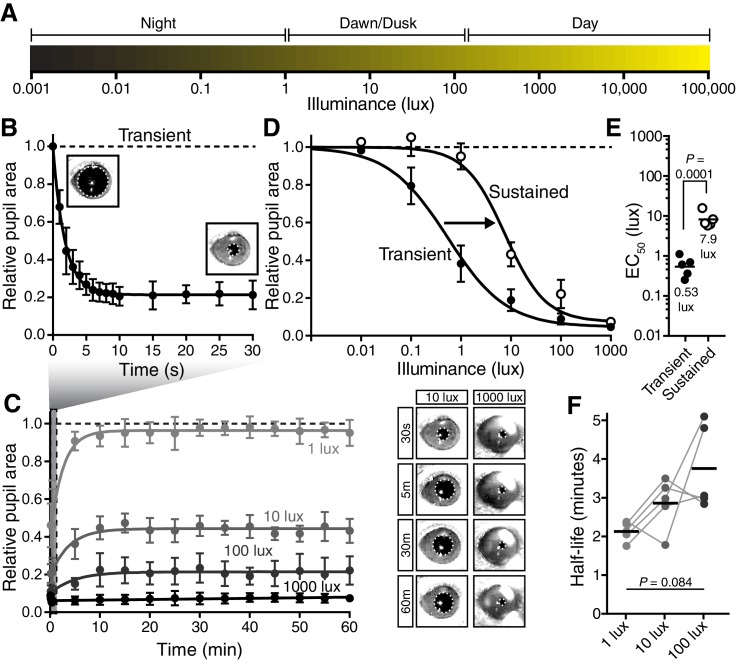
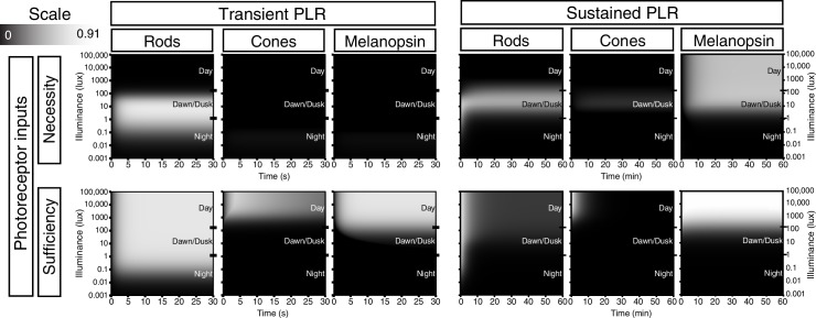
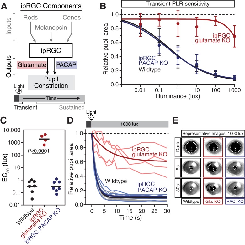
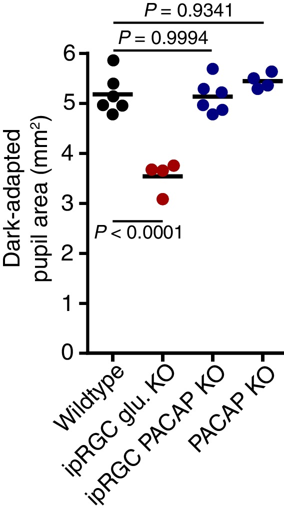
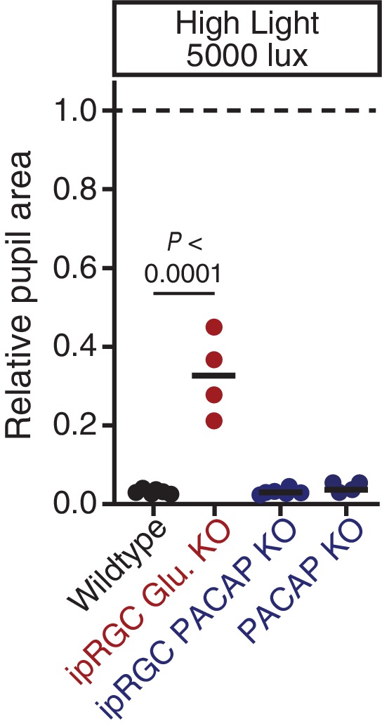
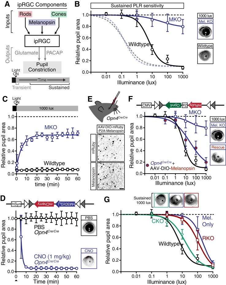
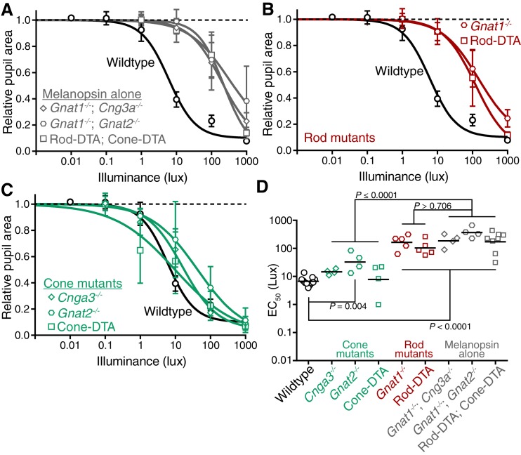
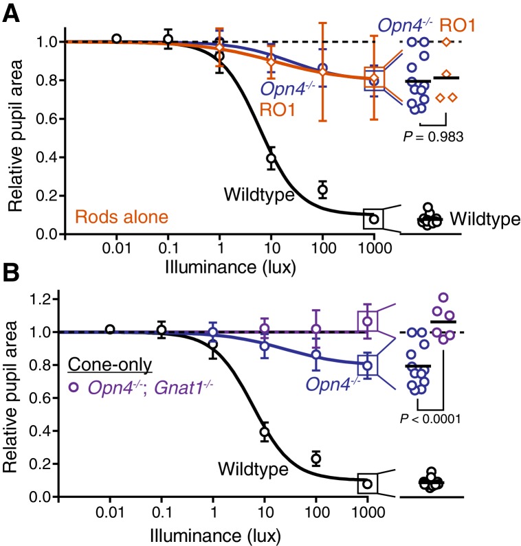
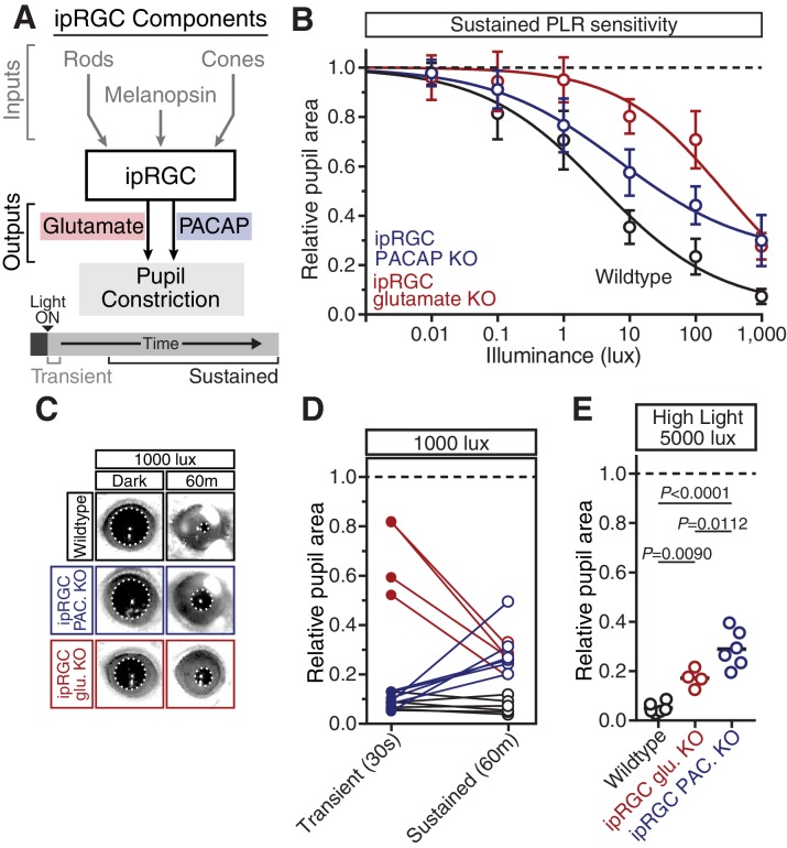
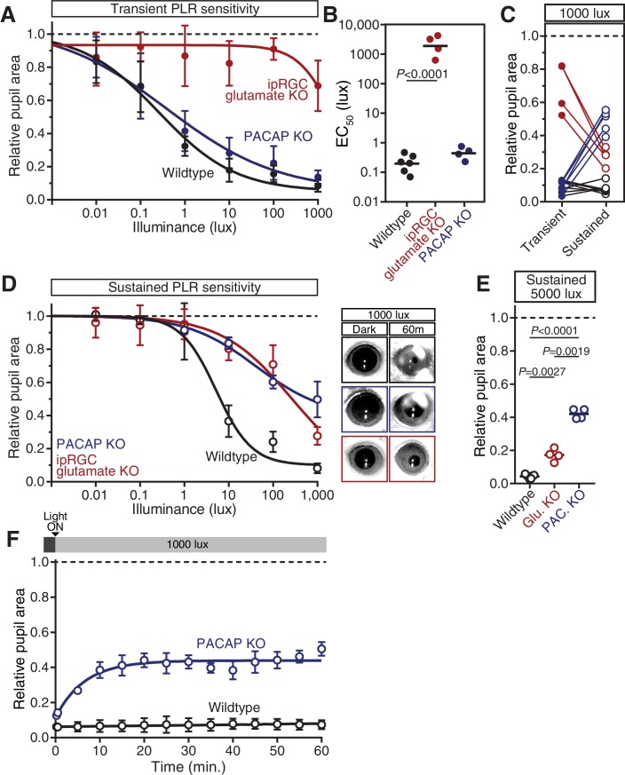
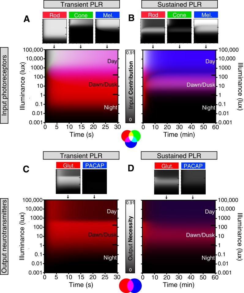
Rapid and stable control of pupil size in response to light is critical for vision, but the neural coding mechanisms remain unclear. Here, we investigated the neural basis of pupil control by monitoring pupil size across time while manipulating each photoreceptor input or neurotransmitter output of intrinsically photosensitive retinal ganglion cells (ipRGCs), a critical relay in the control of pupil size. We show that transient and sustained pupil responses are mediated by distinct photoreceptors and neurotransmitters. Transient responses utilize input from rod photoreceptors and output by the classical neurotransmitter glutamate, but adapt within minutes. In contrast, sustained responses are dominated by non-conventional signaling mechanisms: melanopsin phototransduction in ipRGCs and output by the neuropeptide PACAP, which provide stable pupil maintenance across the day. These results highlight a temporal switch in the coding mechanisms of a neural circuit to support proper behavioral dynamics.

摘要

对光作出快速且稳定的瞳孔大小控制对视觉至关重要,但神经编码机制仍不清楚。在此,我们通过在操纵内在光敏性视网膜神经节细胞(ipRGCs)的每个光感受器输入或神经递质输出时跨时间监测瞳孔大小,来研究瞳孔控制的神经基础,ipRGCs是瞳孔大小控制中的关键中继。我们表明,瞬态和持续的瞳孔反应由不同的光感受器和神经递质介导。瞬态反应利用视杆光感受器的输入,并通过经典神经递质谷氨酸输出,但在数分钟内会适应。相比之下,持续反应由非常规信号机制主导:ipRGCs中的黑视蛋白光转导以及神经肽垂体腺苷酸环化酶激活肽(PACAP)的输出,这在一整天内提供稳定的瞳孔维持。这些结果突出了神经回路编码机制中的一种时间转换,以支持适当的行为动态。

https://cdn.ncbi.nlm.nih.gov/pmc/blobs/1e8c/5079752/cf4de9904399/elife-15392-fig6-figsupp1.jpg
https://cdn.ncbi.nlm.nih.gov/pmc/blobs/1e8c/5079752/f3f3938a6aec/elife-15392-fig1.jpg
https://cdn.ncbi.nlm.nih.gov/pmc/blobs/1e8c/5079752/d93cc9586e0c/elife-15392-fig1-figsupp1.jpg
https://cdn.ncbi.nlm.nih.gov/pmc/blobs/1e8c/5079752/791ce8ef6be1/elife-15392-fig1-figsupp2.jpg
https://cdn.ncbi.nlm.nih.gov/pmc/blobs/1e8c/5079752/8140adcbc207/elife-15392-fig2.jpg
https://cdn.ncbi.nlm.nih.gov/pmc/blobs/1e8c/5079752/bea35550404a/elife-15392-fig2-figsupp1.jpg
https://cdn.ncbi.nlm.nih.gov/pmc/blobs/1e8c/5079752/1e2ce794fe4e/elife-15392-fig2-figsupp2.jpg
https://cdn.ncbi.nlm.nih.gov/pmc/blobs/1e8c/5079752/475da2a0edac/elife-15392-fig2-figsupp3.jpg
https://cdn.ncbi.nlm.nih.gov/pmc/blobs/1e8c/5079752/c3552f5510ea/elife-15392-fig2-figsupp4.jpg
https://cdn.ncbi.nlm.nih.gov/pmc/blobs/1e8c/5079752/6a4936f23048/elife-15392-fig2-figsupp5.jpg
https://cdn.ncbi.nlm.nih.gov/pmc/blobs/1e8c/5079752/378e05f53a65/elife-15392-fig3.jpg
https://cdn.ncbi.nlm.nih.gov/pmc/blobs/1e8c/5079752/222c89c33fca/elife-15392-fig3-figsupp1.jpg
https://cdn.ncbi.nlm.nih.gov/pmc/blobs/1e8c/5079752/e393d7e574a8/elife-15392-fig3-figsupp2.jpg
https://cdn.ncbi.nlm.nih.gov/pmc/blobs/1e8c/5079752/f7c77a861a0e/elife-15392-fig3-figsupp3.jpg
https://cdn.ncbi.nlm.nih.gov/pmc/blobs/1e8c/5079752/8281a7235ee8/elife-15392-fig4.jpg
https://cdn.ncbi.nlm.nih.gov/pmc/blobs/1e8c/5079752/4f6d15f5950e/elife-15392-fig4-figsupp1.jpg
https://cdn.ncbi.nlm.nih.gov/pmc/blobs/1e8c/5079752/3036da578e83/elife-15392-fig4-figsupp2.jpg
https://cdn.ncbi.nlm.nih.gov/pmc/blobs/1e8c/5079752/116450e696b4/elife-15392-fig4-figsupp3.jpg
https://cdn.ncbi.nlm.nih.gov/pmc/blobs/1e8c/5079752/33fc019fd777/elife-15392-fig4-figsupp4.jpg
https://cdn.ncbi.nlm.nih.gov/pmc/blobs/1e8c/5079752/6997414eb025/elife-15392-fig5.jpg
https://cdn.ncbi.nlm.nih.gov/pmc/blobs/1e8c/5079752/b4235abec872/elife-15392-fig5-figsupp1.jpg
https://cdn.ncbi.nlm.nih.gov/pmc/blobs/1e8c/5079752/91cbae895bdd/elife-15392-fig6.jpg
https://cdn.ncbi.nlm.nih.gov/pmc/blobs/1e8c/5079752/cf4de9904399/elife-15392-fig6-figsupp1.jpg

相似文献

1
A visual circuit uses complementary mechanisms to support transient and sustained pupil constriction.视觉回路利用互补机制来支持瞬态和持续性瞳孔收缩。
Elife. 2016 Sep 26;5:e15392. doi: 10.7554/eLife.15392.
2
Temporal characteristics of melanopsin inputs to the human pupil light reflex.黑视蛋白输入到人类瞳孔光反射的时间特征。
Vision Res. 2015 Feb;107:58-66. doi: 10.1016/j.visres.2014.12.001. Epub 2014 Dec 10.
3
A five-primary photostimulator suitable for studying intrinsically photosensitive retinal ganglion cell functions in humans.一种适用于研究人类内在光敏视网膜神经节细胞功能的五原色光刺激器。
J Vis. 2015 Jan 26;15(1):15.1.27. doi: 10.1167/15.1.27.
4
Intrinsically photosensitive retinal ganglion cells.内在光敏性视网膜神经节细胞
J Neuroophthalmol. 2007 Sep;27(3):195-204. doi: 10.1097/WNO.0b013e31814b1df9.
5
Melanopsin and Cone Photoreceptor Inputs to the Afferent Pupil Light Response.黑素视蛋白和视锥光感受器向传入性瞳孔光反应的输入。
Front Neurol. 2019 May 22;10:529. doi: 10.3389/fneur.2019.00529. eCollection 2019.
6
Melanopsin cells are the principal conduits for rod-cone input to non-image-forming vision.黑视蛋白细胞是视杆视锥细胞向非成像视觉输入信息的主要通道。
Nature. 2008 May 1;453(7191):102-5. doi: 10.1038/nature06829. Epub 2008 Apr 23.
7
Melanopsin-mediated post-illumination pupil response in the peripheral retina.外周视网膜中黑视蛋白介导的光照后瞳孔反应。
J Vis. 2016 Jun 1;16(8):5. doi: 10.1167/16.8.5.
8
Melanopsin Cell Dysfunction is Involved in Sleep Disruption in Parkinson's Disease.黑视素细胞功能障碍与帕金森病睡眠障碍有关。
J Parkinsons Dis. 2020;10(4):1467-1476. doi: 10.3233/JPD-202178.
9
[Pupil and melanopsin photoreception].[瞳孔与黑视蛋白光感受]
Nippon Ganka Gakkai Zasshi. 2013 Mar;117(3):246-68; discussion 269.
10
Non-image-forming vision as measured through ipRGC-mediated pupil constriction is not modulated by covert visual attention.通过 ipRGC 介导的瞳孔收缩测量的非成像视觉不受隐蔽视觉注意的调节。
Cereb Cortex. 2024 Mar 1;34(3). doi: 10.1093/cercor/bhae107.

引用本文的文献

1
A stress-sensing circuit signals to the central pacemaker to reprogram circadian rhythms.一个压力感应电路向中央起搏器发出信号,以重新设定昼夜节律。
Sci Adv. 2025 Jun 20;11(25):eadr7960. doi: 10.1126/sciadv.adr7960.
2
A genome-wide in vivo CRISPR screen identifies neuroprotective strategies in the mouse and human retina.全基因组体内CRISPR筛选确定了小鼠和人类视网膜中的神经保护策略。
bioRxiv. 2025 Mar 24:2025.03.22.644712. doi: 10.1101/2025.03.22.644712.
3
Blue Light Damages Retinal Ganglion Cells Via Endoplasmic Reticulum Stress and Autophagy in Chickens.

本文引用的文献

1
Regulation of AMPA receptor phosphorylation by the neuropeptide PACAP38.神经肽PACAP38对AMPA受体磷酸化的调节
Proc Natl Acad Sci U S A. 2015 May 26;112(21):6712-7. doi: 10.1073/pnas.1507229112. Epub 2015 May 11.
2
Photoreceptor regulation of spatial visual behavior.空间视觉行为的光感受器调节
Invest Ophthalmol Vis Sci. 2015 Feb 10;56(3):1842-9. doi: 10.1167/iovs.14-15644.
3
Impaired circadian photosensitivity in mice lacking glutamate transmission from retinal melanopsin cells.缺乏视网膜黑素视蛋白细胞谷氨酸传递的小鼠昼夜节律光敏感性受损。
蓝光通过内质网应激和自噬损伤鸡的视网膜神经节细胞。
Invest Ophthalmol Vis Sci. 2025 Jan 2;66(1):3. doi: 10.1167/iovs.66.1.3.
4
Developmental control of rod number via a light-dependent retrograde pathway from intrinsically photosensitive retinal ganglion cells.通过光依赖性逆行途径从内在光敏性视网膜神经节细胞控制棒的数量。
Dev Cell. 2024 Nov 4;59(21):2897-2911.e6. doi: 10.1016/j.devcel.2024.07.018. Epub 2024 Aug 13.
5
Defining spatial nonuniformities of all ipRGC types using an improved Opn4 recombinase mouse line.利用改良的 Opn4 重组酶小鼠品系定义所有 ipRGC 类型的空间非均一性。
Cell Rep Methods. 2024 Aug 19;4(8):100837. doi: 10.1016/j.crmeth.2024.100837. Epub 2024 Aug 9.
6
Diversity and Molecular Evolution of Nonvisual Opsin Genes across Environmental, Developmental, and Morphological Adaptations in Frogs.非视觉视蛋白基因在青蛙的环境、发育和形态适应中的多样性和分子进化。
Mol Biol Evol. 2024 Jun 1;41(6). doi: 10.1093/molbev/msae090.
7
A pupillary contrast response in mice and humans: Neural mechanisms and visual functions.小鼠和人类的瞳孔对比反应:神经机制与视觉功能
Neuron. 2024 Jul 17;112(14):2404-2422.e9. doi: 10.1016/j.neuron.2024.04.012. Epub 2024 May 1.
8
A new recombinase mouse line to target intrinsically photosensitive retinal ganglion cells (ipRGCs).一种用于靶向内在光敏性视网膜神经节细胞(ipRGCs)的新型重组酶小鼠品系。
bioRxiv. 2024 Apr 20:2024.04.16.589750. doi: 10.1101/2024.04.16.589750.
9
Intercellular communication atlas reveals Oprm1 as a neuroprotective factor for retinal ganglion cells.细胞间通讯图谱揭示 Oprm1 是视网膜神经节细胞的神经保护因子。
Nat Commun. 2024 Mar 11;15(1):2206. doi: 10.1038/s41467-024-46428-z.
10
Sympathetic NPY controls glucose homeostasis, cold tolerance, and cardiovascular functions in mice.交感神经 NPY 控制小鼠的葡萄糖稳态、耐寒性和心血管功能。
Cell Rep. 2024 Feb 27;43(2):113674. doi: 10.1016/j.celrep.2024.113674. Epub 2024 Jan 17.
J Biol Rhythms. 2015 Feb;30(1):35-41. doi: 10.1177/0748730414561545. Epub 2014 Dec 15.
4
Retinal output changes qualitatively with every change in ambient illuminance.视网膜输出会随着环境光照度的每一次变化而发生质的改变。
Nat Neurosci. 2015 Jan;18(1):66-74. doi: 10.1038/nn.3891. Epub 2014 Dec 8.
5
Mice deficient of glutamatergic signaling from intrinsically photosensitive retinal ganglion cells exhibit abnormal circadian photoentrainment.来自内在光敏性视网膜神经节细胞的谷氨酸能信号传导缺陷的小鼠表现出异常的昼夜节律光调节。
PLoS One. 2014 Oct 30;9(10):e111449. doi: 10.1371/journal.pone.0111449. eCollection 2014.
6
Opponent melanopsin and S-cone signals in the human pupillary light response.人眼瞳孔光反射中的拮抗黑素视蛋白和S视锥信号。
Proc Natl Acad Sci U S A. 2014 Oct 28;111(43):15568-72. doi: 10.1073/pnas.1400942111. Epub 2014 Oct 13.
7
A role for melanopsin in alpha retinal ganglion cells and contrast detection.黑视蛋白在α视网膜神经节细胞和对比检测中的作用。
Neuron. 2014 May 21;82(4):781-8. doi: 10.1016/j.neuron.2014.03.022.
8
Dual-transmitter neurons: functional implications of co-release and co-transmission.双递质神经元:共同释放与共同传递的功能意义
Curr Opin Neurobiol. 2014 Dec;29:25-32. doi: 10.1016/j.conb.2014.04.010. Epub 2014 May 13.
9
Effects of exposure to intermittent versus continuous red light on human circadian rhythms, melatonin suppression, and pupillary constriction.间歇性红光与持续红光照射对人体昼夜节律、褪黑素抑制及瞳孔收缩的影响。
PLoS One. 2014 May 5;9(5):e96532. doi: 10.1371/journal.pone.0096532. eCollection 2014.
10
Photoresponse diversity among the five types of intrinsically photosensitive retinal ganglion cells.五种内在光敏感视网膜神经节细胞的光反应多样性。
J Physiol. 2014 Apr 1;592(7):1619-36. doi: 10.1113/jphysiol.2013.262782. Epub 2014 Jan 6.