Kabeer Farha Arakkaveettil, Rajalekshmi Dhanya Sethumadhavan, Nair Mangalam Sivasankaran, Prathapan Remani
Division of Cancer Research, Regional Cancer Centre (RCC), Thiruvananthapuram, India.
Chemical Sciences and Technology Division, CSIR-National Institute for Interdisciplinary Science and Technology, Thiruvananthapuram, India.
Integr Med Res. 2017 Jun;6(2):190-206. doi: 10.1016/j.imr.2017.03.004. Epub 2017 May 31.
Deoxyelephantopin (DOE) is a natural bioactive sesquiterpene lactone from , a traditionally relevant herb in Chinese and Indian medicine. It has shown promising anticancer effects against a broad spectrum of cancers.
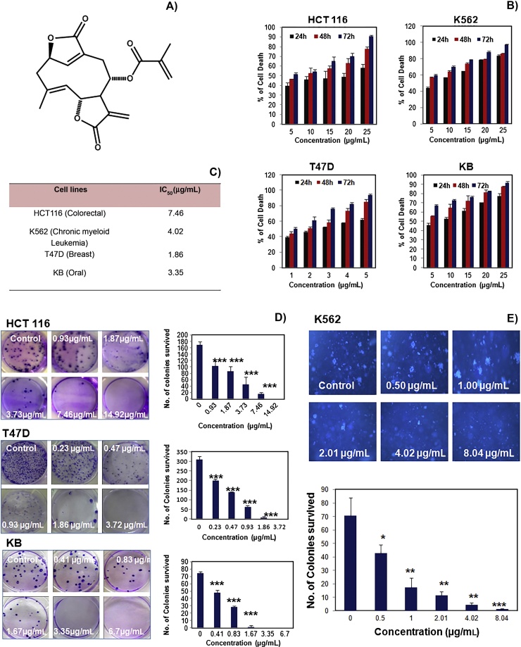
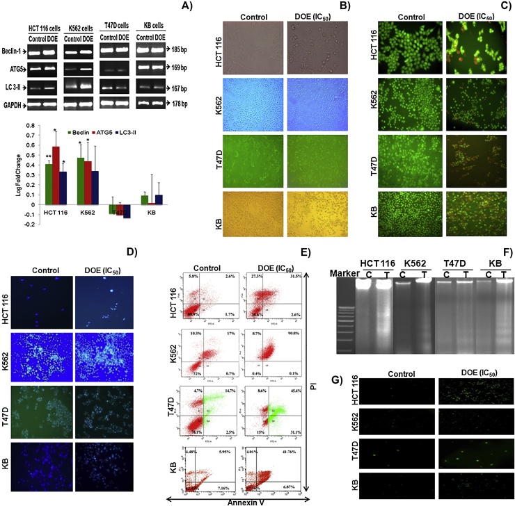
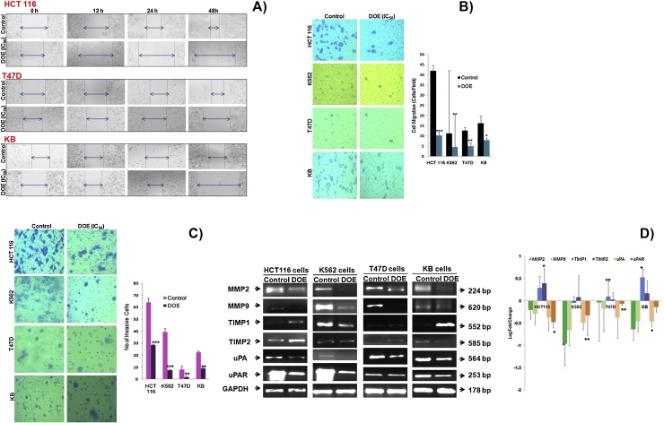
We examined the effect of DOE on growth, autophagy, apoptosis, cell cycle progression, metastasis, and various molecular signaling pathways in cancer cells, and endeavored to decipher the molecular mechanisms underlying its effect. The cytotoxicity of DOE was examined by MTT (3-[4,5-dimethylthiazol-2-yl]-2,5-diphenyltetrazolium bromide) and colony formation assays. The antimetastatic potential of DOE was identified by wound closure, as well as invasion and migration assays. The expression of mRNAs and proteins related to cytotoxicity in cancer cells induced by DOE was investigated using reverse transcription-polymerase chain reaction, flow cytometry, and Western blot analysis.
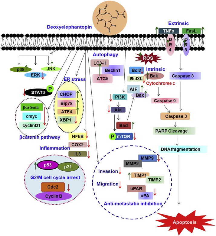
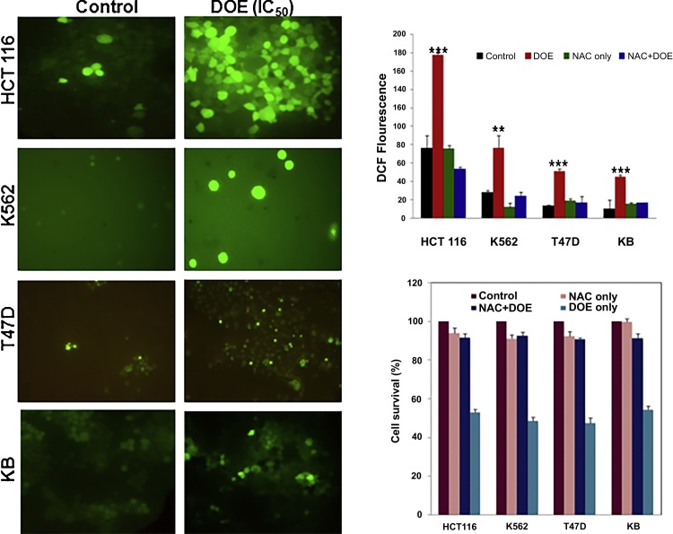
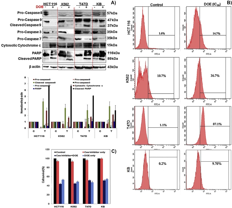
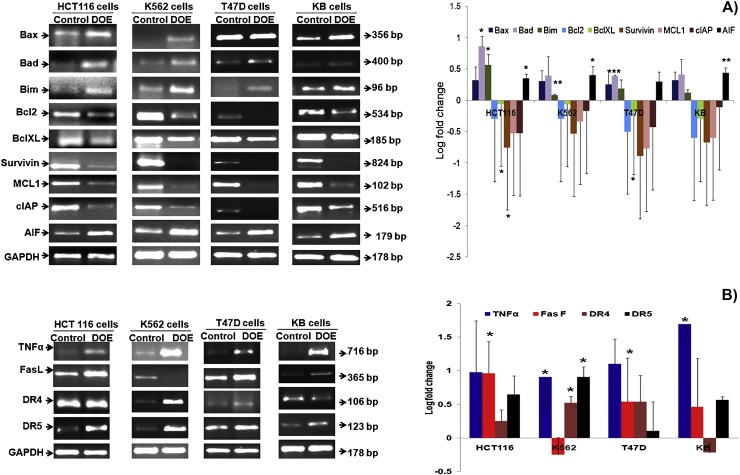
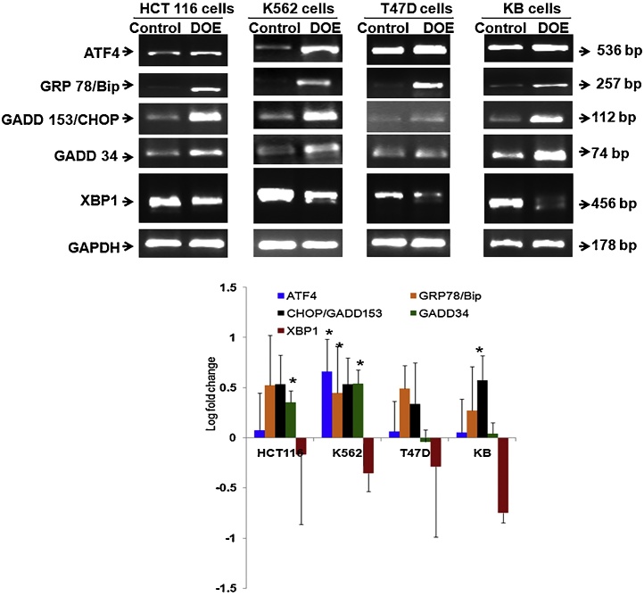
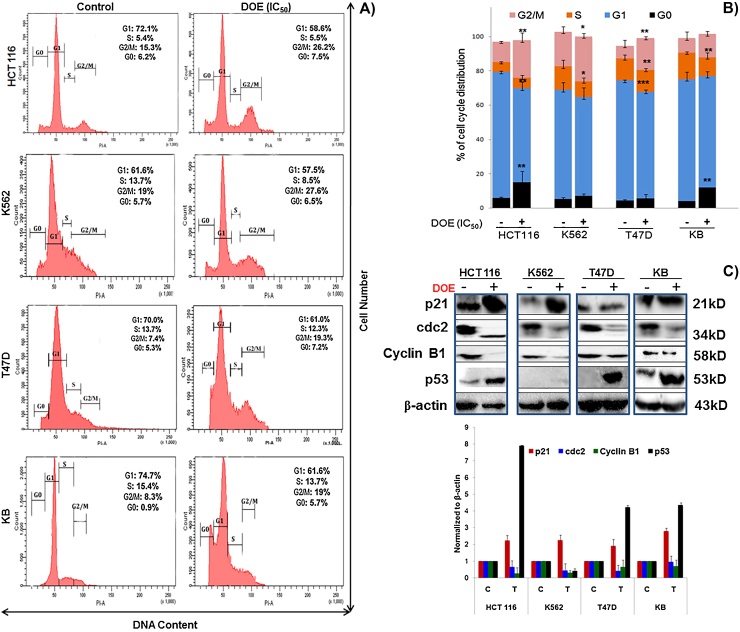
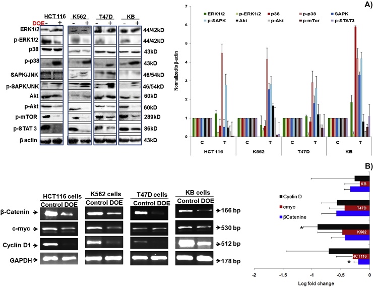
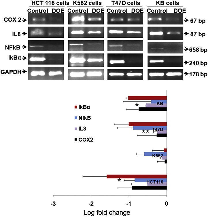
DOE showed significant cytotoxicity and induced apoptosis in cancer cells. DOE promoted the autophagy of HCT 116 and K562 cells. DOE arrested cell cycle progression in the G2/M phase. DOE treatment caused activation of caspase-8, -9, -3 and -7, reactive oxygen species production, and cleavage of cleavage of poly-ADP-ribose polymerase (PARP), the markers of apoptosis. Moreover, apoptosis induction was associated with mitochondrial permeability and endoplasmic reticulum stress. Treatment of cancer cells with DOE inhibited mitogen-activated protein kinases, nuclear factor-kappa B, phosphatidylinositol 3-kinase (PI3K/Akt), and β-catenin signaling. Furthermore, treatment of DOE increased the expression of p53, phospho-Jun amino-terminal kinases (p-JNK), and p-p38 and decreased the expression of phospho-signal transducer and activator of transcription 3 (p-STAT3) and phospho-mammalian target of rapamycin (p-mTOR) in cancer cells. DOE downregulated matrix metalloproteinase (MMP-2) and MMP-9, urokinase-type plasminogen activator (uPA), and urokinase-type plasminogen activator receptor (uPAR) mRNA levels in cancer cells.
These findings concluded that DOE may be useful as a chemotherapeutic agent against cancer.
脱氧地胆草素(DOE)是一种天然生物活性倍半萜内酯,源自一种在中国和印度传统医学中具有重要意义的草药。它已显示出对多种癌症具有有前景的抗癌作用。
我们研究了DOE对癌细胞生长、自噬、凋亡、细胞周期进程、转移及各种分子信号通路的影响,并试图阐明其作用的分子机制。通过MTT(3-[4,5-二甲基噻唑-2-基]-2,5-二苯基四氮唑溴盐)和集落形成试验检测DOE的细胞毒性。通过伤口愈合试验以及侵袭和迁移试验确定DOE的抗转移潜力。使用逆转录-聚合酶链反应、流式细胞术和蛋白质免疫印迹分析研究DOE诱导的癌细胞中与细胞毒性相关的mRNA和蛋白质的表达。
DOE在癌细胞中显示出显著的细胞毒性并诱导凋亡。DOE促进HCT 116和K562细胞的自噬。DOE使细胞周期进程停滞在G2/M期。DOE处理导致半胱天冬酶-8、-9、-3和-7激活、活性氧生成以及聚ADP核糖聚合酶(PARP)裂解,这些都是凋亡的标志物。此外,凋亡诱导与线粒体通透性和内质网应激相关。用DOE处理癌细胞可抑制丝裂原活化蛋白激酶、核因子-κB、磷脂酰肌醇3-激酶(PI3K/Akt)和β-连环蛋白信号通路。此外,DOE处理增加了癌细胞中p53、磷酸化c-Jun氨基末端激酶(p-JNK)和p-p38的表达,并降低了磷酸化信号转导和转录激活因子3(p-STAT3)和磷酸化雷帕霉素靶蛋白(p-mTOR)的表达。DOE下调癌细胞中基质金属蛋白酶(MMP-2)和MMP-9、尿激酶型纤溶酶原激活剂(uPA)和尿激酶型纤溶酶原激活剂受体(uPAR)的mRNA水平。
这些发现表明DOE可能作为一种抗癌化疗药物具有应用价值。